

















How ligaments tear in a sprained ankle and why ankles re-injure
Ankle ligaments can tear when the foot turns inward under load, and repeat sprains can occur when joint stability or movement control remains impaired (Hertel, 2002; Herzog et al., 2019). In lateral sprains, excessive rearfoot supination can strain the supporting ligament complex (Hertel, 2002). The resulting symptoms can settle before joint stability and movement control have fully recovered, and earlier sprains are associated with later injuries (Herzog et al., 2019).
The outer ligaments, the grade of tissue damage and the distinction between sprains and fractures shape the clinical picture. Recovery studies, a rehabilitation trial and reviews of chronic ankle instability show why pain relief, ligament healing and a secure return to activity are separate outcomes. The evidence gives no single recovery timetable.

Evidence last checked 17 Nov 2026 · 14 min
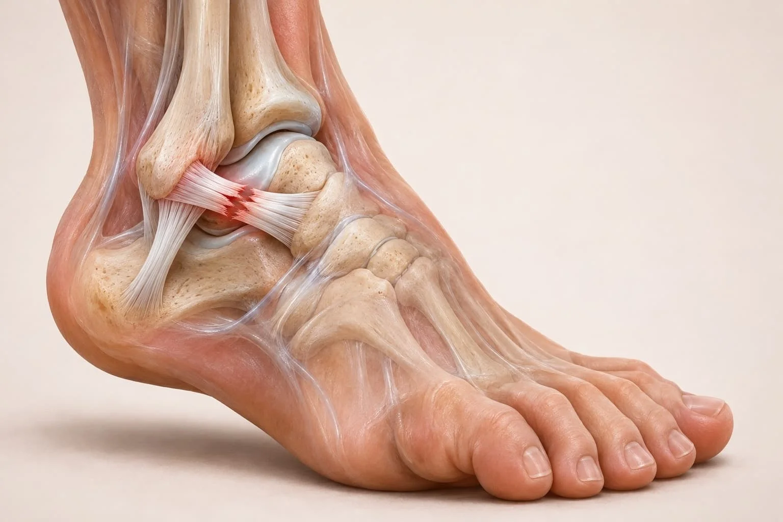
What a sprained ankle is and which ligaments are affected
A ligament connects one bone to another and limits excessive movement at a joint. A sprained ankle occurs when that supporting tissue is stretched or torn, as described by the US National Library of Medicine's MedlinePlus service (2025). It is a tissue injury. The name does not establish which ligament is damaged or whether the disruption is partial or complete.
The distribution of ankle injuries is uneven. In a 2014 systematic review, Doherty and colleagues examined prospective epidemiological studies and identified lateral ankle sprain as the most commonly observed pattern. That finding distinguishes the usual location from the less frequent medial and syndesmotic injuries, without assuming that every painful ankle follows the same mechanism (Herzog et al., 2019).
Lateral ankle ligaments
Three ligaments form the lateral complex. Hertel (2002) identified the anterior talofibular ligament, calcaneofibular ligament and posterior talofibular ligament as restraints across the outer ankle, while the deltoid ligament provides support on the medial side. The anterior talofibular ligament, or ATFL, is particularly exposed to forces that pull the talus forwards and turn it inward.
The calcaneofibular ligament crosses towards the heel bone. It restrains movement at the ankle and subtalar joints. Hertel (2002) identified it as the second most frequently injured outer ligament and described its role in resisting excessive supination. The posterior band is rarely damaged.
Medial and high ankle sprains
The medial or deltoid ligament complex supports the inner ankle; the lower tibiofibular connection forms the syndesmosis. Hertel (2002) placed these structures in different parts of the ankle's ligament system, each contributing to stability under a different pattern of movement. A high ankle sprain involves the syndesmotic region rather than simply the outer bands. The clinical distinction is recognised in the review by Herzog and colleagues (2019).
The distinction matters when describing research findings. Dubin and colleagues (2011) reviewed lateral and syndesmotic sprains separately because their anatomy, injury mechanisms and assessment differ. Data about an ordinary lateral sprain cannot automatically supply a prognosis for a high sprain, particularly when the latter involves the connection between the lower-leg bones. The location must be established before the studies are compared.
Sprained ankle symptoms and grades of ligament damage
MedlinePlus (2025) lists pain, swelling, bruising and inability to move the joint among sprain symptoms. They describe an injured region without revealing the precise fibres that have failed. An ankle can also be painful when the damage includes a fracture or another structure, so symptoms alone do not settle the diagnosis.
Pain, swelling and loss of function
Swelling accompanies local tissue damage. Dubin and colleagues (2011) described bleeding into the space around a ruptured ligament, followed by increased vessel permeability and the movement of inflammatory cells into injured tissue. The visible enlargement of the ankle therefore reflects a biological response as well as the disruption of the ligament itself. Pain may restrict how the foot bears weight.
The 2021 clinical guideline by Martin and colleagues described significant swelling, pain, limited weight bearing and altered walking as features seen after an acute lateral ankle sprain. Those findings guide clinical assessment, but the extent of swelling does not identify a named ligament in isolation. A clinical examination considers both the symptoms and the way the joint behaves under movement and loading.
The symptoms have a variable course. In the 2008 systematic review by van Rijn and colleagues, reports of pain fell rapidly during the first 2 weeks among adults with acute lateral ankle sprains. Yet persistent complaints were recorded at later follow-up. Relief from early pain is therefore a different observation from the absence of longer-term symptoms.
Mild, partial and complete ligament tears
Severity grades refer to the tissue injury and resulting stability. Petersen and colleagues (2013) described grade I as ligament stretching without a macroscopic rupture or joint instability, while grade II involves a partial tear with pain, swelling and some functional limitation. The ligament may remain continuous in the milder pattern. Its capacity to restrain movement can nevertheless be altered.
Grade III describes a complete rupture associated with marked functional impairment and instability (Petersen et al., 2013). That is a structural distinction, not a measure of how dramatic an ankle looks to an observer. Grading also leaves room for injuries of other tissues around the joint, which a ligament label alone cannot account for.
A sprain and a strain describe different tissues. MedlinePlus (2025) defines a strain as injury to a muscle or tendon, while a sprain concerns a ligament. The similarity of pain, swelling and movement restriction can blur these terms in ordinary conversation. Their anatomical definitions remain distinct.
What causes a sprained ankle and how ligaments tear
The injury is commonly generated when the foot turns inward while the ankle is in plantar flexion. Petersen and colleagues (2013) identified this combination of inversion, adduction and plantar flexion as a usual mechanism of lateral ligament injury. It creates strain across stabilising fibres. Whether those fibres stretch or rupture depends on the resulting tissue damage rather than the label applied to the movement.
Inversion and rotational forces
The ankle complex does not function as a single hinge. Hertel (2002) described lateral sprains as excessive rearfoot supination against an externally rotated lower leg, involving the talocrural and subtalar joints. The bony articulations and ligaments constrain movement together. If the joint exceeds the available restraint, tension reaches the supporting bands.
The ATFL has a particular mechanical role. In the biomechanical literature reviewed by Hertel (2002), it limited forward displacement of the talus as well as excessive inversion and internal rotation; tension rose as the ankle moved towards plantar flexion. The CFL contributes a different restraint against excessive supination at the adjacent joints.
Mechanisms differ with location. Herzog and colleagues (2019) separated lateral, medial and syndesmotic injuries in their epidemiological review, while Dubin and colleagues (2011) treated syndesmotic loading as a separate diagnostic problem. A twisting event therefore supplies useful context, but its direction alone does not provide a complete map of the damage.
Ligament fibre disruption
Ligament fibres fail under load. Petersen and colleagues (2013) distinguished stretching without visible rupture from partial fibre disruption and complete tearing. A complete tear may also disturb the restraint provided by the injured joint complex. The clinical consequence is instability rather than simply local tenderness.
Torn connective tissue triggers a second process. Dubin and colleagues (2011) described bleeding near separated ligament ends and the movement of plasma proteins and inflammatory cells into the damaged region. This reaction produces swelling while tissue clearance and repair begin. Local symptoms can therefore persist after the twisting force has ended.
Re-injury cannot be understood from the torn fibres alone. Hertel (2002) described later mechanical changes, altered joint motion and impaired sensorimotor function as components of chronic ankle instability. A joint may have repaired tissue and still respond differently when balance is challenged.
How sprained ankles are diagnosed and fractures distinguished
Clinical assessment seeks to establish ligament damage while keeping possible fractures in view. The 2021 guideline by Martin and colleagues describes history-taking, physical examination and specific stability tests for lateral ankle sprain. Swelling and restricted movement are part of that assessment. The examination must also distinguish symptoms caused by damaged bone from those produced by ligament injury.
Clinical examination and stability tests
The anterior drawer test examines forward movement of the talus relative to the lower leg. Martin and colleagues (2021) included the traditional anterior drawer, reverse anterolateral drawer and anterolateral talar palpation among the clinical tests used to assist diagnosis.
Timing can change diagnostic performance. The 2018 guideline by Vuurberg and colleagues reported 84% sensitivity and 96% specificity for assessment of anterior talofibular damage when the anterior drawer examination was delayed for 4–5 days after injury. Those figures concern that particular examination and timing. They cannot be assigned to a patient assessed at another stage or treated as a general accuracy estimate for all ankle injuries.
Martin and colleagues (2021) also described repeated measurement of swelling, range of movement, talar translation, inversion and balance across an episode of clinical care. Each measure captures a different part of function. A person may regain an acceptable range of motion while still reporting instability under movement, which makes a single examination item insufficient to describe the full recovery course.
Radiography and ligament imaging
Plain radiographs answer a question about bone. Bachmann and colleagues (2003) analysed the Ottawa ankle rules as a method of excluding ankle and midfoot fractures; their meta-analysis included 27 studies involving 15 581 patients. A negative result lowered the likelihood of fracture in the studied clinical settings.
The Ottawa rules have high sensitivity but more limited specificity. Bachmann and colleagues (2003) found that the clinical decision tool could reduce unnecessary radiographs, while retaining its purpose as a means of excluding fractures rather than identifying the type of sprain. Persistent tenderness, clinical concern and the structure of the injury still belong to the wider diagnostic assessment.
Ultrasound addresses the soft tissues directly. In a 2024 systematic review, Kocsis and colleagues evaluated ultrasound against MRI using eight studies and 434 patients with acute lateral ligament injuries. The authors concluded that ultrasound had high diagnostic accuracy for acute lateral ligament damage in the included studies. Operator-dependent examination and the type of ligament assessed remain relevant when applying those results to a particular injury.
MRI can reveal associated damage that a routine ankle examination may leave uncertain. The 2018 guideline by Vuurberg and colleagues discussed its use when high-grade ligament, syndesmotic, osteochondral or occult fracture injury is suspected. The scope is selective. The presence of a sprain alone is not evidence that every injured ankle requires the same imaging examination.
How sprained ankles heal and what recovery research shows
Ligament repair and symptom recovery follow related but different courses. The systematic review by Petersen and colleagues (2013) described inflammatory, proliferative and remodelling phases, with substantial variation in duration between individuals. Tissue repair is therefore a process rather than a single point at which all functions return simultaneously. A visible reduction in swelling cannot establish the tensile properties of the repaired fibres.
Ligament healing and remodelling
After disruption, inflammatory cells enter the damaged area while local blood vessels respond. Dubin and colleagues (2011) described the formation of a haematoma around torn ligament ends, followed by granulation tissue and the activity of fibroblasts during repair. Fibroblasts deposit collagen. The initial material bridges the injured region before its organisation changes under subsequent remodelling.
The collagen fibres gradually align and develop cross-links during the remodelling phase, according to Dubin and colleagues (2011). Petersen and colleagues (2013) placed inflammatory, proliferative and maturation changes within a longer model of ligament healing, while specifying that the phases vary from person to person. The model explains why repaired tissue can evolve after initial pain has fallen.
Clinical follow-up tests add a second view of healing. Petersen and colleagues (2013) described collagen formation during the proliferative phase and stated that controlled ligament stress promotes collagen fibre orientation. Such descriptions should be distinguished from direct measurements of a patient's strength. A review of clinical outcomes is needed to judge whether movement restrictions and symptoms have resolved.
Functional rehabilitation and recovery outcomes
In their 2008 review, van Rijn and colleagues included 31 studies of adults with acute lateral ankle sprains, of which 24 were judged high quality. Pain reports fell rapidly within the first 2 weeks. The speed of early symptom reduction did not translate into a uniform longer-term result. Across the studies, 5%–33% still reported pain after 1 year.
Reported full recovery was also variable. The review by van Rijn and colleagues (2008) found that 36%–85% of patients described full recovery within a period of 3 years, with different study populations and follow-up methods. These percentages are ranges of results from different studies. They do not form a single estimate for an individual, and the endpoint of full recovery was not necessarily identical across investigators.
The 2021 practice guideline by Martin and colleagues concluded that supervised exercise directed at identified impairments could support recovery and reduce reinjury. Petersen and colleagues (2013) also reviewed functional treatment compared with longer immobilisation, while finding that conclusions were sensitive to the quality of the trials included. These results describe studied care pathways. They are not instructions for the treatment of an individual sprain.
Return to an activity is a narrower outcome than the disappearance of symptoms. Martin and colleagues (2021) distinguished return to participation from full recovery without limitations, which can take substantially longer and may not occur in all patients. Research into physical activity recommendations addresses the broader value of movement, while ankle-sprain studies concern injury-specific recovery. Both subjects involve activity, but their outcomes are different.
Why ankle sprains recur and chronic instability develops
A previous ankle sprain is associated with subsequent injury, but recurrent sprains do not all share one explanation. Herzog and colleagues (2019) reviewed the relationship between a first sprain, later sprains and adverse long-term outcomes such as chronic ankle instability. Repeated episodes may coexist with ligament laxity or altered movement control. The mechanisms can overlap in the same ankle.
Mechanical ligament laxity
Mechanical instability refers to changes in how the joint is physically restrained. Hertel (2002) listed pathological laxity, altered joint mechanics, synovial irritation and degenerative changes among the possible mechanical contributors to chronic ankle instability. These abnormalities are not interchangeable. Laxity concerns restraint under load, whereas irritation and structural degeneration describe different tissue states.
Complete ligament disruption may affect mechanical restraint directly. Petersen and colleagues (2013) distinguished a fully ruptured ligament with instability from a mild sprain without macroscopic rupture or unstable joint movement. The distinction does not predict recovery by grade alone. Long-term function also depends on structures and movement systems outside the original torn fibres (Hertel, 2002).
Balance, proprioception and movement control
Functional instability concerns control rather than merely the passive tightness of the joint. Hertel (2002) attributed it to deficiencies in proprioception and neuromuscular control, while noting that mechanical and functional instability may occur together. Proprioception refers to the information used to detect joint position and movement. A joint that feels unreliable can therefore present a different problem from demonstrable ligament laxity.
The 2021 guideline by Martin and colleagues considered sensorimotor performance and dynamic postural control in the assessment of chronic ankle instability. Balance, range of motion and functional movement can vary independently of reported pain.
The return to sport also takes place within wider athletic demands. Sport nutrition and athletic performance concerns energy and performance, whereas the ankle studies by Hupperets and colleagues (2009) measured recurrent sprains after an injury. A measure of sporting capacity cannot substitute for the injury outcome. The original trial judged recurrence by athletes' reports.
Findings from recurrence studies
Lin and colleagues (2021) reviewed nine studies involving 3804 physically active participants aged 15–32, including students, soldiers and athletes. Chronic ankle instability prevalence was 25%, with study estimates ranging from 7% to 53%; among participants with a history of ankle sprain, the reported prevalence was 46%, ranging from 9% to 76%. All participants were physically active.
A direct intervention comparison adds information about recurrence. In a 2009 randomised trial, Hupperets and colleagues assigned 522 athletes with a previous lateral sprain to usual care or usual care with additional proprioceptive training. During the one-year follow-up, 56 athletes in the additional-training group reported recurrence, compared with 89 controls, corresponding to the reported proportions of 22% and 33%. The main endpoint was self-reported reinjury.
Study | Population or design | Outcome | Result |
Lin et al., 2021 | Nine studies; 3804 active participants | Chronic ankle instability prevalence | 25%; range 7%–53% |
van Rijn et al., 2008 | Review of adults with acute lateral sprains | Re-sprain across varying follow-up periods | 3%–34% |
Hupperets et al., 2009 | Randomised trial; 522 athletes; one-year follow-up | Self-reported repeat ankle sprain | 22% with extra training; 33% with usual care |
Across all participants, additional proprioceptive training reduced the risk of self-reported recurrent ankle sprains (RR 0.63, 95% CI 0.45–0.88). The researchers also reported separate subgroup results according to whether athletes had received medical treatment for their initial injury.
Among athletes who received medical treatment for their initial ankle sprain, the intervention did not significantly reduce self-reported recurrences or recurrences leading to lost sports time. However, a statistically significant reduction was observed for recurrences associated with healthcare or productivity costs (RR 0.24, 95% CI 0.08–0.72).
What is proven about sprained ankle recovery and recurrence
The anatomical mechanism is established: lateral ligaments restrain ankle motion and can be damaged by excessive inversion-related loading (Hertel, 2002; Petersen et al., 2013). Clinical evidence also supports the distinction between early pain relief and complete functional recovery. The review by van Rijn and colleagues (2008) found wide variation in later pain, recovery and reinjury reports.
A sprained ligament may heal while movement control or joint stability remains impaired. A previous sprain is associated with recurrence, but the size of that risk depends on the studied population and outcome (Herzog et al., 2019; Hertel, 2002).
There is direct trial evidence that repeat sprains can be reduced in a particular athletic setting. Hupperets and colleagues (2009) found fewer self-reported recurrent injuries following additional proprioceptive training than following usual care alone. The finding concerns athletes and the programme studied. It cannot supply a universal recurrence percentage for everyone with an ankle sprain.
Risk prediction remains more uncertain. Vuurberg and colleagues (2018) reported that a pooled association between previous lateral ankle sprain and a subsequent sprain was not statistically significant, with a risk ratio of 1.44 and a 95% confidence interval of 0.96–2.16. That result sits alongside the associations and intervention findings described in other studies. The difference reflects questions, populations and methods that are not identical.
Long-term prevalence is particularly sensitive to definition. Lin and colleagues (2021) found a wide spread of chronic instability estimates and identified differences in study exclusion criteria, age and sport as possible explanations. Prospective follow-up with a consistent definition would give clearer estimates of which injuries lead to lasting instability. A single recovery time or a single percentage cannot describe all ligament grades and activity settings.
Frequently asked questions
How long does a sprained ankle take to heal?
Recovery time varies with injury severity and what is counted as recovery. The review by van Rijn and colleagues (2008) found an early reduction in pain but continuing symptoms in some patients after a year. Tissue remodelling can continue after initial improvement (Petersen et al., 2013). Return to activity and complete recovery are distinct outcomes.
Can you walk on a sprained ankle?
Weight bearing remains possible for some people with a sprain. Martin and colleagues (2021) included limited weight bearing and altered gait among findings after acute lateral sprain. Walking ability alone does not identify the grade of a tear or exclude damage to bone, which requires separate assessment.
What is the difference between a sprained ankle and a fracture?
A sprain damages a ligament connecting bones at a joint, whereas a fracture damages bone (MedlinePlus, 2025; Bachmann et al., 2003). Both can cause pain and restrict loading. The Ottawa ankle rules assess fracture likelihood; Bachmann and colleagues reviewed their diagnostic accuracy. Ligament integrity is a different question from fracture screening.
Is a sprain different from a strain?
Yes. A sprain involves the fibrous ligament tissue connecting bones, while a strain involves a muscle or tendon, according to MedlinePlus (2025). Both injuries can produce swelling and pain. The terms distinguish the structure injured; neither term by itself states how severe the damage is or whether another tissue is also injured.
Can an ankle sprain heal completely?
Complete recovery is possible, but it is not universal in the studies reviewed. The review by van Rijn and colleagues (2008) reported full recovery in 36%–85% of patients within 3 years across different studies. Martin and colleagues (2021) distinguished returning to participation from the absence of symptoms or limitation. Residual instability can remain after an initial sprain.
Sources
- Herzog MM, Kerr ZY, Marshall SW, Wikstrom EA. Epidemiology of Ankle Sprains and Chronic Ankle Instability. Journal of Athletic Training. 2019;54(6):603–610. DOI 10.4085/1062-6050-447-17.
- Lin CI, Houtenbos S, Lu YH, Mayer F, Wippert PM. The epidemiology of chronic ankle instability with perceived ankle instability- a systematic review. Journal of Foot and Ankle Research. 2021;14:41. DOI 10.1186/s13047-021-00480-w.
- Doherty C, Delahunt E, Caulfield B, et al. The incidence and prevalence of ankle sprain injury: a systematic review and meta-analysis of prospective epidemiological studies. Sports Medicine. 2014;44:123–140. DOI 10.1007/s40279-013-0102-5.
- Hertel J. Functional Anatomy, Pathomechanics, and Pathophysiology of Lateral Ankle Instability. Journal of Athletic Training. 2002;37(4):364–375.
- Martin RL, Davenport TE, Fraser JJ, et al. Ankle Stability and Movement Coordination Impairments: Lateral Ankle Ligament Sprains Revision 2021. Journal of Orthopaedic & Sports Physical Therapy. 2021;51(4):CPG1–CPG80. DOI 10.2519/jospt.2021.0302.
- Hupperets MDW, Verhagen EALM, van Mechelen W. Effect of unsupervised home based proprioceptive training on recurrences of ankle sprain: randomised controlled trial. BMJ. 2009;339:b2684. DOI 10.1136/bmj.b2684.
- van Rijn RM, van Os AG, Bernsen RMD, et al. What is the clinical course of acute ankle sprains? A systematic literature review. The American Journal of Medicine. 2008;121(4):324–331.e6. DOI 10.1016/j.amjmed.2007.11.018.
- Petersen W, Rembitzki IV, Koppenburg AG, et al. Treatment of acute ankle ligament injuries: a systematic review. Archives of Orthopaedic and Trauma Surgery. 2013;133:1129–1141. DOI 10.1007/s00402-013-1742-5.
- Dubin JC, Comeau D, McClelland RI, Dubin RA, Ferrel E. Lateral and syndesmotic ankle sprain injuries: a narrative literature review. Journal of Chiropractic Medicine. 2011;10(3):204–219. DOI 10.1016/j.jcm.2011.02.001.
- Bachmann LM, Kolb E, Koller MT, Steurer J, ter Riet G. Accuracy of Ottawa ankle rules to exclude fractures of the ankle and mid-foot: systematic review. BMJ. 2003;326(7386):417. DOI 10.1136/bmj.326.7386.417.
- Kocsis K, Stubnya B, Váncsa S, et al. Diagnostic accuracy of ultrasonography in acute lateral ankle ligament injury: a systematic review and meta-analysis. Injury. 2024;55(Suppl 3):111730. DOI 10.1016/j.injury.2024.111730.
- Vuurberg G, Hoorntje A, Wink LM, et al. Diagnosis, treatment and prevention of ankle sprains: update of an evidence-based clinical guideline. British Journal of Sports Medicine. 2018;52(15):956. DOI 10.1136/bjsports-2017-098106.
- MedlinePlus, US National Library of Medicine. Sprains and Strains. Last updated 22 September 2025. https://medlineplus.gov/sprainsandstrains.html

Ellen Marsh — author
Ellen writes Health & Medicine explainers for Cabdirect, with a focus on public health, disease prevention and the interpretation of medical research for general readers. Her academic background combines biomedical sciences with postgraduate study in populatio...
This article is for general information and is not medical advice. It was reviewed for accuracy by a qualified clinician; decisions about your health should be made with your own doctor.
Related

WHO 2010 Physical Activity Recommendations by Age Group

Sport Nutrition: Energy, Fuel and Athletic Performance

How exercise affects self-concept and well-being in midlife

How self-talk patterns relate to competitive anxiety in sport

Visceral fat and why exercise shrinks the fat stored around your organs

Dehydration symptoms and how fluid loss during exercise affects the body

Piriformis syndrome and how a hip muscle irritates the sciatic nerve