














How Graves’ disease antibodies push the thyroid into overdrive
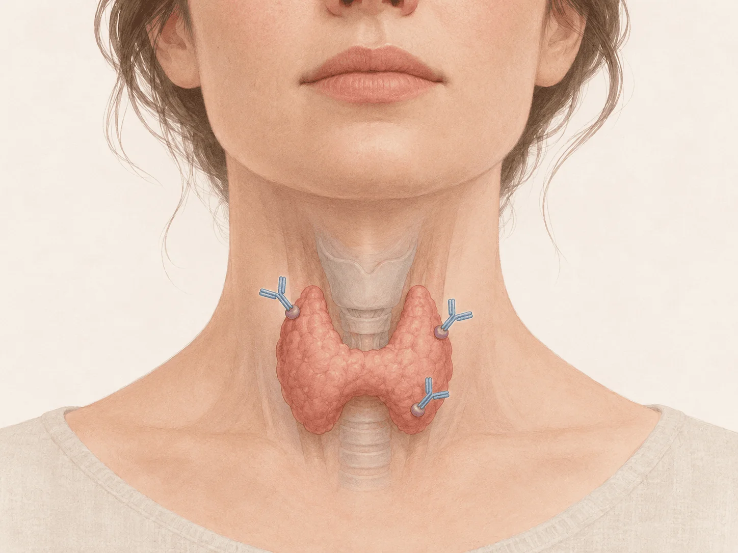
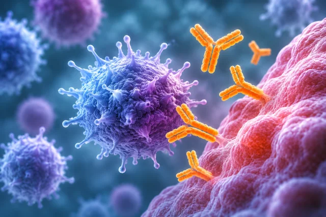
Graves’ disease antibodies push the thyroid into overdrive by activating the same receptor that normally responds to pituitary thyroid-stimulating hormone, so the gland keeps making and releasing thyroid hormone even after normal feedback has suppressed TSH. The key antibodies are usually described as TSH-receptor antibodies, or TRAb, and the stimulating fraction is often called thyroid-stimulating immunoglobulin, or TSI.
Key points include the receptor-level mechanism, the link to hyperthyroidism and goitre, systemic effects, extra-thyroidal eye and skin disease, and diagnostic testing. The evidence base includes major endocrine reviews, professional guidelines and medical information from national and academic organisations.

Evidence last checked 10 Nov 2026 · 12 min
What makes Graves disease an autoimmune form of hyperthyroidism
Graves’ disease is an [autoimmune disorder](/health?sub=vaccines-immunity) in which the immune system loses tolerance to the thyroid-stimulating hormone receptor, or TSHR. The receptor sits on the surface of thyroid follicular cells and normally converts a pituitary signal into intracellular activity that increases hormone synthesis, hormone release and thyroid-cell growth. Reviews by Smith and Hegedüs (2016), Davies et al. (2020) and Lanzolla et al. (2024) identify TSHR autoantibodies as the central pathogenic feature that separates Graves’ hyperthyroidism from many other causes of an overactive thyroid.
Under normal physiology, circulating thyroid hormones restrain the system through negative feedback. When thyroxine and triiodothyronine rise, the pituitary releases less TSH, reducing the signal to the thyroid. In Graves’ disease, stimulating autoantibodies bypass that control: they continue to activate TSHR even when the pituitary has appropriately lowered TSH, so a low blood TSH does not mean the thyroid is receiving no stimulation.
The term TRAb is an umbrella label rather than a guarantee that every antibody has the same effect. Laboratory and immunology reviews distinguish stimulating antibodies, blocking antibodies and antibodies with little direct functional effect; Graves’ hyperthyroidism is driven primarily by the stimulating group (Gao et al., 2024; Kalra et al., 2024). TSI and thyroid-stimulating antibody are commonly used for assays or antibody activity that specifically reflects stimulation, while some TRAb binding assays detect antibodies that occupy the receptor without directly showing what they do to it.
The receptor itself is a useful clue to why the disease is so persistent. TSHR is designed to translate an external signal into repeated intracellular responses, so an antibody that behaves like an agonist can exploit the same signalling machinery as the natural hormone. The pituitary can reduce its own TSH output, but it has no comparable feedback switch that directly turns off antibody production. That separation between endocrine feedback and immune activity is central to understanding why biochemical hyperthyroidism can continue despite a very low TSH concentration (Davies et al., 2020; Smith and Hegedüs, 2016).
How do Graves disease antibodies keep the thyroid switched on
When a stimulating Graves’ antibody binds TSHR, it behaves as a receptor agonist. The receptor activates signalling pathways inside the thyroid cell, particularly the cyclic-AMP pathway, increasing the machinery used to take up iodine, build thyroid hormone and release it into the circulation. The same persistent signal promotes thyroid-cell growth, which helps explain why Graves’ disease often produces a diffuse goitre rather than a single overactive nodule (Davies et al., 2020; Kahaly et al., 2018).
This is why the disease can look paradoxical on a laboratory panel. Blood TSH is usually suppressed because the pituitary senses too much circulating thyroid hormone, yet the thyroid remains active because antibodies are providing a separate receptor signal. NIDDK (2021) describes TSI as acting like TSH, while the American Thyroid Association (accessed 2026) similarly explains that TRAb or TSI bind to receptors on thyroid cells and stimulate excess thyroid hormone production.
The stimulation is not simply a one-time trigger. Autoantibody production is sustained by an autoimmune process involving T cells, B cells and failure of immune tolerance, so the gland can remain overactive until the autoimmune drive changes or treatment alters thyroid hormone production or thyroid tissue. No single initiating event explains all cases; current reviews describe genetic susceptibility interacting with environmental and physiological influences rather than a universal trigger (Lanzolla et al., 2024).
Thyroid-related signalling also has roles outside human thyroid disease. For example, [research on seasonal reproduction in temperate birds](/animals/seasonal-reproduction-temperate-birds-animals) describes how thyroid-hormone pathways contribute to seasonal reproductive timing. That mechanism is distinct from the antibody-driven thyroid stimulation seen in Graves’ disease.
Graves’ hyperthyroidism is not caused by excess pituitary TSH. It is caused by autoantibodies that keep the TSH receptor switched on after normal feedback has already suppressed TSH.
Why does excess thyroid hormone cause symptoms across the body
Thyroid hormones regulate energy use and influence the cardiovascular, nervous, gastrointestinal, musculoskeletal and reproductive systems. Once stimulating antibodies have driven hormone production above the body’s needs, tissues throughout the body are exposed to a stronger thyroid-hormone signal. That broad reach is why Graves’ disease can produce a mixture of heat intolerance, sweating, tremor, sleep disturbance, weight loss, frequent bowel movements, muscle weakness and a rapid or irregular heartbeat rather than one local thyroid symptom (De Leo et al., 2016; NIDDK, 2021).
Many of the most noticeable manifestations are consequences of hyperthyroidism rather than direct antibody attack on each organ. Increased adrenergic responsiveness contributes to palpitations, tremor and nervousness; accelerated metabolic activity contributes to heat intolerance and weight loss; and prolonged excess thyroid hormone can increase bone turnover. NIDDK (2021) describes cardiovascular complications, including rapid or irregular heartbeat, as well as thinning bones and osteoporosis among the potential consequences of untreated Graves’ disease.
The clinical picture does not map perfectly onto a single hormone concentration. Age, baseline health, duration of hormone excess and individual tissue sensitivity all influence how strongly symptoms are felt. This matters when interpreting Graves’ disease as a systemic condition: the autoantibody starts the thyroid problem, but the resulting hormone excess then becomes the main driver of many symptoms and complications.
Cardiovascular and skeletal effects illustrate that distinction. Persistent thyroid-hormone excess increases heart rate, contractility and susceptibility to rhythm disturbance, while chronically increased bone turnover can reduce bone density over time. These downstream effects are clinically important even though the antibodies are not directly attacking the heart or skeleton. In other words, Graves’ disease begins as an autoimmune receptor disorder in the thyroid, then produces a broader endocrine state whose consequences depend on how much hormone is produced and how long tissues are exposed to it (NIDDK, 2021; De Leo et al., 2016).
Common hyperthyroid effects described by official clinical sources include:
- fast or irregular heartbeat and increased awareness of the heartbeat
- heat intolerance, sweating and warm skin
- tremor, nervousness, irritability or disturbed sleep
- weight loss despite normal or increased appetite
- more frequent bowel movements and changes in muscle strength
Why can Graves disease affect the eyes and skin as well as the thyroid
Graves’ orbitopathy, also called thyroid eye disease, is not simply the eyeball reacting to high thyroid hormone. The immune process can also involve cells in the orbit, especially fibroblasts that express TSHR and interact with the insulin-like growth factor 1 receptor. Immune signalling can drive inflammation, glycosaminoglycan accumulation, tissue swelling and changes in orbital fat and extraocular muscles, producing symptoms such as grittiness, redness, lid retraction, proptosis and double vision (Bartalena et al., 2021; Lanzolla et al., 2024).
Because the eye process has its own local immune biology, its timing and severity are not locked to the blood thyroid-hormone level. Eye symptoms may appear before, around the time of, or after recognition of thyroid dysfunction, and some people with thyroid eye disease can have relatively modest biochemical thyroid abnormalities. EUGOGO guidance also treats high TSHR-antibody concentrations as one risk marker for Graves’ orbitopathy, supporting a link between the autoimmune response and the extra-thyroidal disease (Bartalena et al., 2021).
Orbital fibroblasts can respond by producing large amounts of hydrophilic extracellular matrix, which draws water into the tissues, and some fibroblast populations can shift towards fat-cell differentiation. Together with inflammatory-cell infiltration and enlargement of extraocular muscles, these changes help explain why the orbit can become crowded and why the eyes may protrude or move less freely. The modern model therefore links antibody-related receptor signalling to a local tissue response rather than treating eye disease as a simple complication of high T4 or T3 alone (Bartalena et al., 2021; Lanzolla et al., 2024).
Estimates of eye involvement vary because study populations and diagnostic definitions differ. De Leo et al. (2016) reported Graves’ orbitopathy in about 25% of people with Graves’ disease, while a meta-analysis by Ippolito et al. (2021) estimated an overall prevalence of about 30%, with an estimate of 25% in studies conducted after 2000. NIDDK (2021) states that more than one in three people with Graves’ disease develop Graves’ ophthalmopathy. These figures reflect differences in populations, periods and definitions rather than a single universal prevalence.
A much less common extra-thyroidal manifestation is Graves’ dermopathy, or pretibial myxoedema, in which skin becomes thickened and textured, usually over the shins or feet. NIDDK (2021) describes Graves’ dermopathy, or pretibial myxoedema, as a rare manifestation that usually affects the shins and can also involve the tops of the feet or other areas. The shared theme is immune activation in tissues capable of responding to the same or related signalling network, not merely exposure to excess circulating thyroid hormone.
Reported frequency of selected extra-thyroidal manifestations
Source | Outcome described | Reported frequency | Context |
De Leo et al., 2016 | Graves’ orbitopathy | About 25% | Peer-reviewed review of hyperthyroidism and Graves’ disease |
Ippolito et al., 2021 | Graves’ orbitopathy | About 30% overall; 25% in post-2000 studies | Meta-analysis of newly diagnosed Graves’ disease |
NIDDK, 2021 | Graves’ ophthalmopathy | More than 1 in 3 | National institute patient information; most cases described as mild |
NIDDK, 2021 | Graves’ dermopathy / pretibial myxoedema | Rare | Usually affects the shins; may also involve the feet or other areas |
How do blood tests show that Graves disease is the cause
Diagnosis has two linked questions: whether hyperthyroidism is present and whether Graves’ autoimmunity is causing it. Thyroid-function testing typically shows suppressed TSH with elevated free T4, T3, or both, establishing biochemical thyrotoxicosis. Antibody testing then provides a more disease-specific clue: a positive TRAb result in the right clinical setting supports Graves’ disease by showing the presence of antibodies directed against the TSH receptor, while a positive TSI or TSAb functional assay additionally demonstrates stimulating activity at that receptor (Kahaly et al., 2018).
Professional guidance recognises TRAb measurement as a useful way to distinguish Graves’ disease from other causes of thyrotoxicosis when the cause is not already clear. The 2016 American Thyroid Association guideline lists TRAb, radioactive iodine uptake and thyroid blood-flow assessment by ultrasonography as diagnostic approaches that can establish aetiology depending on the clinical setting and local expertise. The 2018 European Thyroid Association guideline likewise describes TSHR-antibody measurement as a sensitive and specific tool for rapid diagnosis and differential diagnosis.
Assay names can be confusing because a binding test and a functional bioassay answer slightly different laboratory questions. Binding assays detect immunoglobulins that interact with TSHR, whereas functional assays assess whether those antibodies stimulate receptor signalling. Modern reviews emphasise that this distinction is relevant when interpreting the words TRAb, TSI and TSAb, even though all refer to the same receptor-centred autoimmune system (Gao et al., 2024; Kalra et al., 2024).
The importance of defining what an antibody assay detects extends beyond thyroid medicine. A [historical study of cardiolipin in syphilis serology](/health/cardiolipin-standardization-syphilis-serology-health) examined the standardisation of a different type of laboratory test. It illustrates a broader issue in serology, but does not provide evidence about the diagnostic accuracy of Graves’ disease antibody assays.
That distinction also explains why antibody results are interpreted alongside thyroid-function tests rather than in isolation. A binding assay may show that receptor-directed antibodies are present, while a functional assay can indicate stimulating activity, but neither single result replaces the biochemical picture created by TSH, free T4 and T3. Reviews of current laboratory practice emphasise combining the antibody result with the clinical phenotype and thyroid hormone measurements, particularly when the presentation is atypical or when more than one autoimmune antibody activity may be present (Gao et al., 2024; Kalra et al., 2024).
When antibody results do not settle the cause, imaging can provide physiological context rather than directly measuring autoimmunity. Diffusely increased radioactive iodine uptake or increased thyroid blood flow can support Graves’ disease, while other patterns point towards alternative causes of thyrotoxicosis. Ross et al. (2016) and the American Thyroid Association (accessed 2026) describe radioactive iodine uptake testing as an additional diagnostic approach when antibody testing is negative or unavailable, while thyroid blood-flow ultrasonography may be useful in selected cases.
What is established about Graves antibodies, and what remains uncertain
The central mechanism is well established: stimulating TSHR autoantibodies activate the thyroid receptor and drive excessive thyroid hormone production. Multiple independent reviews and guidelines converge on that model, and diagnostic practice uses the same biology by measuring TRAb or TSI. The evidence is therefore strong for the statement that Graves’ hyperthyroidism is receptor-mediated autoimmune stimulation rather than a pituitary command to the thyroid (Smith and Hegedüs, 2016; Davies et al., 2020; Ross et al., 2016; Lanzolla et al., 2024).
Several boundaries remain important. The precise combination of genetic, immune and environmental events that breaks tolerance in an individual is not predictable from routine tests, and an antibody concentration is not a simple meter of symptom severity. A person can have mixed antibody activities, while eye disease can behave partly independently of the biochemical intensity of hyperthyroidism. These uncertainties do not challenge the TSHR mechanism; they explain why Graves’ disease has a variable clinical course.
Evidence about thyroid autoimmunity should not be confused with research on unrelated preventive interventions. For example, [research on selenium and cancer prevention](/health/selenium-for-preventing-cancer-health) addresses cancer outcomes rather than the TSH-receptor antibodies responsible for Graves’ hyperthyroidism. Its findings cannot establish whether selenium changes the autoimmune mechanism of this thyroid disease.
The extra-thyroidal biology is also more complex than the thyroid mechanism. TSHR is implicated in orbital tissues, but current models include interaction with IGF1R, immune-cell signalling, fibroblast activation and tissue remodelling (Bartalena et al., 2021; Lanzolla et al., 2024). Research continues to refine how these pathways connect and why only a subset of people with Graves’ disease develop clinically important eye or skin involvement.
The evidence is strongest for the receptor-to-hormone sequence. Autoimmunity creates receptor-directed antibodies; stimulating antibodies activate TSHR; the thyroid overproduces hormone; hormone excess produces most [systemic symptoms](/health). Variation lies in how the autoimmune response begins, how long it persists, which antibody activities dominate and whether tissues outside the thyroid become involved.
Frequently asked questions
Is Graves disease the same as hyperthyroidism?
No. Hyperthyroidism means the thyroid is producing too much hormone, while Graves’ disease is an autoimmune cause of that state. Graves’ antibodies activate the TSH receptor and drive thyroid overactivity. Other conditions can also cause hyperthyroidism, so the terms describe related but different things.
Why is TSH low in Graves disease if the thyroid is being stimulated?
TSH falls because the pituitary senses high circulating thyroid hormone and reduces its own signal through normal negative feedback. The thyroid remains stimulated because Graves’ antibodies activate the TSH receptor independently of pituitary TSH. This mismatch is a hallmark of the disease mechanism.
Are TSI and TRAb the same thing?
They refer to the same receptor-centred autoimmune process but are not always identical laboratory labels. TRAb is a broad term for antibodies that bind the TSH receptor. TSI and TSAb refer to stimulating TSH-receptor antibodies, but assay formats differ: cell-based bioassays directly measure stimulatory activity, whereas some automated TSI assays detect antibodies by binding rather than by measuring receptor signalling.
Can Graves disease occur without eye symptoms?
Yes. The thyroid is the main target, and many people with Graves’ disease never develop clinically important thyroid eye disease. De Leo et al. (2016) reported Graves’ orbitopathy in about one quarter of affected people, while NIDDK (2021) reports it in more than one third. Eye involvement is therefore common but not required.
Can Graves antibodies be present when thyroid hormone levels are normal?
They can. Antibody activity and thyroid hormone levels are related but do not move in perfect lockstep, especially across different phases of disease or after treatment has altered thyroid function. Antibody results therefore need to be interpreted alongside TSH, thyroid hormones, symptoms and the broader clinical context.
Sources
- Davies TF, Andersen S, Latif R, et al. Graves’ disease. Nature Reviews Disease Primers. 2020;6:52. DOI: 10.1038/s41572-020-0184-y.
- Smith TJ, Hegedüs L. Graves' Disease. New England Journal of Medicine. 2016;375(16):1552–1565. DOI: 10.1056/NEJMra1510030.
- Ross DS, Burch HB, Cooper DS, et al. 2016 American Thyroid Association Guidelines for Diagnosis and Management of Hyperthyroidism and Other Causes of Thyrotoxicosis. Thyroid. 2016;26(10):1343–1421. DOI: 10.1089/thy.2016.0229.
- Kahaly GJ, Bartalena L, Hegedüs L, Leenhardt L, Poppe K, Pearce SH. 2018 European Thyroid Association Guideline for the Management of Graves' Hyperthyroidism. European Thyroid Journal. 2018;7(4):167–186. DOI: 10.1159/000490384.
- Bartalena L, Kahaly GJ, Baldeschi L, et al. The 2021 European Group on Graves' orbitopathy clinical practice guidelines for the medical management of Graves' orbitopathy. European Journal of Endocrinology. 2021;185(4):G43–G67. DOI: 10.1530/EJE-21-0479.
- Lanzolla G, Marinò M, Menconi F. Graves disease: latest understanding of pathogenesis and treatment options. Nature Reviews Endocrinology. 2024;20(11):647–660. DOI: 10.1038/s41574-024-01016-5.
- Gao Y, Qiu L, Yu S, Cheng X. Thyroid stimulating receptor autoantibodies. Clinica Chimica Acta. 2024;559:119700. DOI: 10.1016/j.cca.2024.119700.
- Kalra S, Selim S, Shrestha D, et al. Best practices in the laboratory diagnosis, prognostication, prediction, and monitoring of Graves’ disease: role of TRAbs. BMC Endocrine Disorders. 2024;24:274. DOI: 10.1186/s12902-024-01809-9.
- National Institute of Diabetes and Digestive and Kidney Diseases. Graves’ Disease. Last reviewed November 2021. https://www.niddk.nih.gov/health-information/endocrine-diseases/graves-disease.
- De Leo S, Lee SY, Braverman LE. Hyperthyroidism. The Lancet. 2016;388(10047):906–918. DOI: 10.1016/S0140-6736(16)00278-6.
- Ippolito S, Cusini C, Lasalvia P, et al. Change in newly diagnosed Graves’ disease phenotype between the twentieth and the twenty-first centuries: meta-analysis and meta-regression. Journal of Endocrinological Investigation. 2021;44(8):1707–1718. DOI: 10.1007/s40618-020-01479-z.
- American Thyroid Association. Graves' Disease. Accessed 2 November 2026. https://www.thyroid.org/graves-disease/.

Ellen Marsh — author
Ellen writes Health & Medicine explainers for Cabdirect, with a focus on public health, disease prevention and the interpretation of medical research for general readers. Her academic background combines biomedical sciences with postgraduate study in populatio...
This article is for general information and is not medical advice. It was reviewed for accuracy by a qualified clinician; decisions about your health should be made with your own doctor.
Related

Selenium and cancer prevention: what the evidence shows

How hormones time seasonal reproduction in temperate-zone birds

How Cardiolipin Helped Standardize Syphilis Serology

How the tetanus shot protects and why boosters are needed
Psoriatic arthritis and why a skin condition affects the joints

How myasthenia gravis antibodies block nerve signals to muscles
What autoimmune disease is and why the immune system attacks the body