

















How HPV causes plantar warts on the soles of the feet
A plantar wart develops when human papillomavirus, or HPV, infects epidermal cells on the sole and stimulates localised thickening of the skin. It may form a painful, inward-growing lesion beneath a weight-bearing area, but the appearance and course differ among lesions.
Clinical descriptions, viral genotyping studies and school-based cohorts explain the relationship between HPV and foot warts. Trials comparing wart treatments add a separate measure of clearance. The evidence distinguishes characteristic changes in skin and virus types from predictions about whether an individual wart will disappear or respond to an intervention.

Evidence last checked 17 Nov 2026 · 13 min
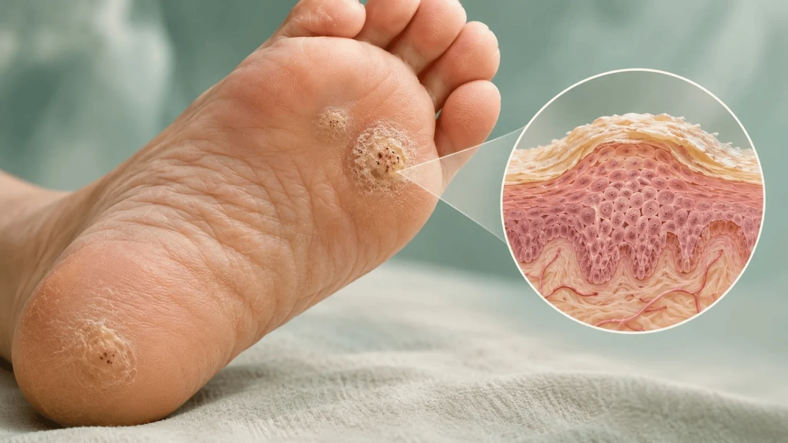
What a plantar wart is and where it develops
A plantar wart is an HPV-associated growth of the skin on the sole of the foot, designated verruca plantaris in dermatological classification (Sterling et al., 2014). The thickened surface reflects changes in infected epidermal cells. Sterling and colleagues (2014) note that corns and calluses can be confused with plantar warts, while Patil and colleagues (2023) documented interrupted skin lines within wart lesions.
Plantar lesions can grow inwards at pressure sites. The 2021 review by Magalhães and colleagues describes hyperkeratotic papules that grow inward at pressure sites and may be deep or superficial. The corresponding variation in tenderness is part of the clinical description, not a measurement of the amount of virus (Magalhães et al., 2021).
Lesions on the weight-bearing sole
At a point of sustained pressure, a plantar wart often grows inward rather than projecting prominently above the surrounding surface (Magalhães et al., 2021). The thickened covering can make it appear embedded in the foot. Some lesions also contain small haemorrhagic points within the hardened skin (Magalhães et al., 2021). This pattern underlies the association between weight-bearing location and pain described in the same review.
An examination of Dutch primary schoolchildren illustrates the range of lesions beyond specialist clinics. In the 2013 cohort of Bruggink and colleagues, 366 of 1,099 participating children had cutaneous warts at baseline, including plantar and other cutaneous lesions, so that proportion is not plantar-wart prevalence alone. The researchers examined feet as well as hands during follow-up (Bruggink et al., 2013).
Solitary and mosaic warts
A plantar wart may occur alone or in a cluster, and the EVerT trial defined a mosaic wart as numerous, closely aggregated plantar warts on the sole (Cockayne et al., 2011). A mosaic is therefore a pattern of neighbouring lesions, rather than a separate virus species. The EVerT trial classified these aggregated lesions separately during analysis (Cockayne et al., 2011).
In a 1982 histopathological study of 50 plantar warts, Laurent and colleagues associated HPV1 with deeper, painful myrmecia and HPV2 with superficial lesions that were often mosaic and painless. The association was reported for the 50 examined warts, not a population-wide sample (Laurent et al., 1982).
Plantar wart symptoms and changes in foot skin
Pain may accompany thickened lesions at weight-bearing sites, as described by Magalhães and colleagues (2021), while Patil and colleagues (2023) observed small red or black dots by dermoscopy in clinically diagnosed warts. Some lesions are superficial and painless in the older HPV typing series (Laurent et al., 1982).
The appearance is not determined by thickness alone. Corns and calluses can also form hardened patches on the feet, and clinical inspection can confuse them with warts (Sterling et al., 2014). Their differences become clearer when structures within the lesion are seen with magnification (Patil et al., 2023).
Black dots and disrupted skin lines
Small black or red points within a wart correspond to vascular or haemorrhagic changes rather than roots. In a 2023 cross-sectional study of 70 clinic patients with palmoplantar warts, corns or calluses, Patil and colleagues found homogeneous black or red dots in all 36 clinically diagnosed wart cases examined by dermoscopy. Magnification also revealed interrupted dermatoglyphics, the normal ridge patterns of palmar and plantar skin.
The darker spots have an anatomical explanation. The British Association of Dermatologists' guideline describes pinpoint bleeding when elongated dermal papillae expose their capillary loops (Sterling et al., 2014). The visible dots therefore arise from structures within the skin and are not evidence of a wart growing long roots into the foot. Their appearance can vary with the overlying keratin.
Pain under pressure and thickened skin
Pain is linked to location and lesion form. The 2021 dermatology review describes deep and superficial plantar papules, with pronounced pain when they occupy pressure areas (Magalhães et al., 2021). A small visible surface can cover a thicker inward-growing area. The review describes this growth pattern, while Laurent and colleagues (1982) associated deep myrmecia with HPV1 in their histological series.
Discomfort is not universal. Laurent and colleagues' 1982 series connected HPV2 with superficial, painless plantar lesions in the warts examined. In a separate school cohort, parents of 73 children reported inconvenience from warts, while 106 reported treatment during follow-up (Bruggink et al., 2013). The parental reports covered cutaneous warts of the hands and feet rather than plantar-specific pain scores (Bruggink et al., 2013).
What causes plantar warts and how HPV spreads
Cutaneous HPV enters skin where small defects expose the basal layer of the epidermis (Magalhães et al., 2021). Contact with infected skin provides one route of transmission. Environmental contact is another route recognised in dermatological guidance, although the length of time infectious HPV survives on a surface is not known (Sterling et al., 2014).
The virus family contains types that favour different tissues. Warts of the hands and soles are associated with cutaneous types, including HPV1, HPV2, HPV27 and HPV57 (Magalhães et al., 2021; de Planell-Mas et al., 2017). Redzic and colleagues (2023) and de Planell-Mas and colleagues (2017) reported different distributions among their sampled lesions.
Skin contact and contaminated surfaces
Direct contact and microscopic skin injury provide a route for HPV infection. A breach in the skin is also involved in staphylococcal skin infections, although these bacterial infections differ from cutaneous HPV. Shared surfaces and footwear were described as possible indirect exposures by Magalhães and colleagues (2021), although the presence of such an exposure does not identify the actual route of infection in an individual case. Laboratory evidence on surface persistence cannot be translated into a precise infectious interval for human HPV (Sterling et al., 2014).
In a 2013 prospective cohort of Dutch schoolchildren, Bruggink and colleagues related new cutaneous warts to household and classroom exposures, with family-wart exposure associated with a hazard ratio of 2.08 (95% CI 1.52–2.86). The observational exposure associations did not directly trace HPV from one person to another (Bruggink et al., 2013).
HPV types associated with plantar lesions
Plantar warts do not arise from one HPV type. Laurent and colleagues (1982) documented HPV1-associated and HPV2-associated lesions with distinct histology in a series of 50 warts. Their findings linked viral identity to appearance within that group. Subsequent molecular studies broadened the set of genotypes detected in plantar samples (de Planell-Mas et al., 2017).
In 2017, de Planell-Mas and colleagues found HPV57 in 37.1%, HPV27 in 23.8%, HPV1a in 20.9% and HPV2 in 15.2% of 105 analysed plantar warts, while Redzic and colleagues (2023) detected HPV27 in 73%, HPV57 in 63% and HPV2 in 42% of persistent cutaneous warts. In Redzic and colleagues' sample, 205 of 269 lesions (76.2%) contained multiple HPV types, so these percentages overlap. Moreover, 98% were older than six months and 92% previously treated; the frequencies mainly describe persistent warts.
How HPV infects the skin and produces plantar warts
The transformation from contact to a visible wart begins in the epidermis. Through minor breaks in skin, HPV reaches basal keratinocytes and follows the differentiation of epidermal cells during replication (Magalhães et al., 2021), while local proliferation produces visible thickening (Sterling et al., 2014).
Viral replication and the visible lump are related but distinct phenomena. Sterling and colleagues (2014) attribute wart formation to infection at the basal epidermal layer followed by local proliferation and hyperkeratinisation. Viral type influences the appearance of the resulting lesion, as the classical HPV1 and HPV2 histological comparison demonstrates (Laurent et al., 1982).
Entry into epidermal keratinocytes
A break in the skin exposes cells in the basal epidermal layer. HPV particles then encounter the keratinocytes that support infection (Magalhães et al., 2021). The review describes attachment to cell-surface structures and movement of viral genetic material towards the nucleus. Viral replication precedes the visible surface growth described by Sterling and colleagues (2014).
The virus relies on the normal movement of epidermal cells towards the surface. Magalhães and colleagues (2021) divide HPV replication into early amplification, maintenance in proliferating cells and later production of viral particles as cells differentiate. The infected area can persist during the maintenance phase. Viral particle production occurs as infected epidermal cells complete their differentiation (Magalhães et al., 2021).
Keratin growth and local immune responses
The main visible consequence is hyperkeratinisation, an accumulation of thickened epidermal material after infection (Sterling et al., 2014). Laurent and colleagues (1982) found different microscopic cell changes in their HPV1 and HPV2 lesions, including enlarged keratohyaline-like granules in myrmecia. The same clinical label can therefore cover lesions with different histological patterns.
Host immunity affects persistence and clearance. Magalhães and colleagues (2021) describe local cytotoxic T cells and helper T cells involved in immune responses to HPV lesions, while noting that complete viral elimination may not occur. Warts can be extensive and difficult to clear in people with impaired cell-mediated immunity (Sterling et al., 2014). Neither study measured an individual time-to-clearance from those immune mechanisms (Magalhães et al., 2021; Sterling et al., 2014).
How plantar warts are diagnosed and distinguished from calluses
Diagnosis usually rests on the characteristic appearance of a wart on the sole. The British Association of Dermatologists describes clinical and histological distinctions between a wart and other thickened skin lesions (Sterling et al., 2014). Dermoscopy, which magnifies surface patterns, can show blood-vessel structures hidden by the thickened outer layer (Patil et al., 2023).
A callus is a different clinical finding, although it may resemble a wart on initial inspection. Sterling and colleagues' 2014 guideline identifies corns and calluses as common diagnostic alternatives on the foot. Patil and colleagues (2023) found different dermoscopic patterns in clinically diagnosed warts, corns and calluses.
Clinical inspection and dermoscopy
In a 2023 cross-sectional study of 70 clinic patients with palmoplantar warts, corns or calluses, Patil and colleagues found homogeneous black or red dots in all 36 clinically diagnosed wart cases, with different patterns in corns and calluses. The wart images also showed interruption of the normal skin lines (Patil et al., 2023).
The findings were not identical across the other diagnoses. A translucent central core was seen in 92.85% of unpared corns and 100% of pared corns; homogeneous opacity appeared in 75% of unpared calluses and 100% of pared calluses (Patil et al., 2023). These proportions describe the examined clinic cases. The study did not measure the diagnostic accuracy of those features in every possible foot lesion.
Biopsy and tests for uncertain lesions
Clinical appearance does not resolve every diagnosis. The 2014 British dermatology guideline explains that warts can be distinguished clinically or histologically from other keratotic lesions, including squamous cell carcinoma (Sterling et al., 2014). Histological assessment can distinguish some lesions, while dermoscopic images in Patil and colleagues (2023) showed interrupted skin markings in clinically diagnosed palmoplantar warts.
Viral genotyping addresses a different issue: which HPV type is present. de Planell-Mas and colleagues (2017) analysed 105 plantar warts for HPV genotypes. In the 105 examined lesions, the molecular analysis identified HPV types at different frequencies (de Planell-Mas et al., 2017).
How plantar warts resolve and what treatment trials show
Warts may persist with little visible inflammation or disappear after an immune response. Sterling and colleagues (2014) describe spontaneous clearance over months or years, whereas a school cohort by Bruggink and colleagues measured children's wart resolution and Redzic and colleagues (2023) examined persistent, previously treated lesions.
Randomised trials test treatment comparisons under defined conditions. In the EVerT trial, there was no statistically significant difference in plantar wart clearance between salicylic acid and cryotherapy at the primary assessment (Cockayne et al., 2011). Kwok and colleagues (2012) reported different site-specific estimates for salicylic acid and no statistically significant difference for cryotherapy against placebo in the foot-wart subgroup.
Spontaneous clearance and recurrence
In a 2013 Dutch prospective cohort, Bruggink and colleagues followed children with cutaneous warts and measured a complete resolution rate of 52 per 100 person-years (95% confidence interval 44–60). When newly developing warts were excluded, the rate for baseline lesions was 90 per 100 person-years, with a 95% confidence interval of 79–100. The rate includes children who received treatment (38% of those with baseline warts); it is neither an untreated clearance rate nor a monthly probability (Bruggink et al., 2013).
Age and wart characteristics also entered the analysis. Younger age and non-Caucasian skin type predicted higher resolution; wart type and size did not.
Wart number was weakly associated with resolution in the adjusted model (HR 1.1 per additional wart; 95% CI 1.0–1.2; P = 0.04) (Bruggink et al., 2013). All cutaneous warts examined in that school population contributed to the estimates. The findings do not provide an adult plantar-wart prognosis.
Persistence and later appearance can have different explanations. The dermatology guideline reports that cell-mediated immune impairment is associated with prolonged wart duration (Sterling et al., 2014). The 2021 HPV review describes the possibility of viral latency after an apparent immune response (Magalhães et al., 2021). Neither description supplies a numerical recurrence probability for plantar warts.
Salicylic acid and cryotherapy trials
Cockayne and colleagues' 2011 EVerT study randomised 240 people aged 12 years and over with plantar warts in podiatry and primary-care settings. Complete clearance after 12 weeks was recorded in 17 of 119 participants (14%) assigned to salicylic acid and 15 of 110 (14%) assigned to cryotherapy, with no statistically significant difference. At six months, self-reported clearance also showed no statistically significant difference between groups (Cockayne et al., 2011).
Kwok and colleagues (2012) reviewed 85 cutaneous-wart trials involving 8,815 randomised participants and found that salicylic acid favoured clearance over placebo among foot-wart cases, with a risk ratio of 1.29 (95% confidence interval 1.07–1.55). The estimate was smaller than that for hand warts, but those subgroup results did not establish how an individual plantar wart would respond.
Study | Participants or lesions | Recorded outcome | Boundary of inference |
Cockayne et al., 2011 | 240 patients with plantar warts | Clearance at 12 weeks: 17/119 (14%) with salicylic acid; 15/110 (14%) with cryotherapy | No statistically significant between-group difference |
Kwok et al., 2012 | 85 trials; 8,815 randomised participants | Salicylic acid versus placebo for foot warts: risk ratio 1.29 (95% CI 1.07–1.55) | A subgroup of a broader cutaneous wart review |
Bruggink et al., 2013 | Children with cutaneous warts | Observed resolution rate: 52 per 100 person-years (95% CI 44–60) | Hand and foot warts; some children received treatment |
Bruggink et al., 2013 | HPV-typed warts in a trial secondary analysis | Wait-and-see clearance at 13 weeks: 21/36 (58%) for HPV1 plantar warts; 4/58 (7%) for HPV2/27/57 plantar warts | Selected genotype groups, not general cure probabilities |
Other interventions and study limitations
Kwok and colleagues' 2012 Cochrane review evaluated other interventions, including topical medicines, injected agents, laser techniques and photodynamic therapy. The authors could not combine results from many of these trials because the interventions and outcomes differed. Some showed possible benefit, while others did not outperform placebo.
HPV genotype may also modify the observed course. At 13 weeks under watchful waiting, Bruggink and colleagues (2013) reported clearance in 21 of 36 (58%) HPV1-associated plantar warts and 4 of 58 (7%) HPV2/27/57-associated plantar warts. These were selected genotype groups within a trial. Laurent and colleagues' (1982) findings connect some lesion appearances with genotype in a small series, but they do not determine treatment response.
What is proven about plantar warts and what is not
Plantar warts are cutaneous HPV infections of the sole that begin in epidermal keratinocytes and can produce inward growth of thickened skin at pressure sites (Sterling et al., 2014; Magalhães et al., 2021). Dermoscopic dark points and interrupted skin lines help distinguish them from corns and calluses in the examined clinical series (Patil et al., 2023). They belong to the broader field of infectious diseases, but diagnosis and treatment findings for plantar warts must be interpreted specifically in relation to cutaneous HPV.
Cutaneous HPV can produce a persistent plantar wart, but neither its appearance nor a treatment label determines when the lesion will clear.
A Dutch prospective study associated new cutaneous warts in schoolchildren with household and classroom exposure to people with warts, although it did not trace the viral source of individual infections (Bruggink et al., 2013). Redzic and colleagues (2023) and de Planell-Mas and colleagues (2017) found different HPV distributions among the lesions in their respective studies.
Treatment claims depend on the comparison and the site. EVerT found no significant difference between salicylic acid and cryotherapy for plantar warts, while the Cochrane review reported a modest advantage for salicylic acid over placebo in foot-wart trials (Cockayne et al., 2011; Kwok et al., 2012). These are distinct research questions, with no contradiction between their findings. At six months, EVerT found no statistically significant difference in self-reported clearance, without reporting a post-clearance recurrence rate in that comparison (Cockayne et al., 2011).
Frequently asked questions
Are plantar warts contagious?
Yes. Cutaneous HPV can move through direct skin contact, and transmission from contaminated surfaces is also described in dermatological research (Magalhães et al., 2021). A Dutch school study associated new cutaneous warts with household and classroom exposure (Bruggink et al., 2013). It did not determine the exact route for each infection.
Can a plantar wart disappear without treatment?
Yes. Spontaneous wart clearance is documented over periods ranging from months to years (Sterling et al., 2014). A Dutch cohort recorded resolution among schoolchildren with cutaneous warts, including lesions on hands and feet (Bruggink et al., 2013). The cohort does not predict when a particular plantar wart will disappear (Bruggink et al., 2013).
How long can a plantar wart last?
A plantar wart may remain for months or years before clearing (Sterling et al., 2014). Duration differs with host immune function, and impaired cell-mediated immunity is associated with longer persistence. The course observed in children with cutaneous warts cannot be treated as the expected duration for an adult with persistent plantar lesions.
Do the black dots in plantar warts mean they have roots?
No. The points reflect small blood vessels or bleeding within a wart, rather than roots extending into the foot (Sterling et al., 2014). Dermoscopy of clinically diagnosed palmoplantar warts in Patil and colleagues' 2023 study found black or red dots together with interrupted skin lines. The dots are a visible diagnostic feature.
Are plantar warts caused by the same HPV types as genital warts?
Usually not. Plantar-wart studies identify cutaneous genotypes including HPV1, HPV2, HPV27 and HPV57 (de Planell-Mas et al., 2017). A dermatology review describes HPV6 and HPV11 as common types in genital HPV lesions (Magalhães et al., 2021). The shared virus-family name does not mean that plantar and genital lesions have the same usual HPV types.
Sources
- Magalhães GM, Vieira EC, Garcia LC, et al. Update on human papilloma virus - part I: epidemiology, pathogenesis, and clinical spectrum. Anais Brasileiros de Dermatologia. 2021;96(1):1–16. DOI 10.1016/j.abd.2020.11.003.
- Laurent R, Kienzler JL, Croissant O, Orth G. Two anatomoclinical types of warts with plantar localization: specific cytopathogenic effects of papillomavirus. Type I (HPV-1) and type 2 (HPV-2). Archives of Dermatological Research. 1982;274(1–2):101–111. DOI 10.1007/BF00510364.
- Sterling JC, Gibbs S, Haque Hussain SS, Mohd Mustapa MF, Handfield-Jones SE. British Association of Dermatologists’ guidelines for the management of cutaneous warts 2014. British Journal of Dermatology. 2014;171(4):696–712. DOI 10.1111/bjd.13310.
- Patil S, Borkar M, Pande S, Meshram K, Oke M. Dermoscopic Findings in Clinically Diagnosed Cases of Plantar Warts, Corns, and Calluses: A Cross-Sectional Study. Cureus. 2023;15(4):e38093. DOI 10.7759/cureus.38093.
- Bruggink SC, Eekhof JAH, Egberts PF, et al. Natural Course of Cutaneous Warts Among Primary Schoolchildren: A Prospective Cohort Study. Annals of Family Medicine. 2013;11(5):437–441. DOI 10.1370/afm.1508.
- Cockayne S, Hewitt C, Hicks K, et al. Cryotherapy versus salicylic acid for the treatment of plantar warts (verrucae): a randomised controlled trial. BMJ. 2011;342:d3271. DOI 10.1136/bmj.d3271.
- Kwok CS, Gibbs S, Bennett C, Holland R, Abbott R. Topical treatments for cutaneous warts. Cochrane Database of Systematic Reviews. 2012;(9):CD001781. DOI 10.1002/14651858.CD001781.pub3.
- Bruggink SC, Eekhof JAH, Egberts PF, et al. Warts transmitted in families and schools: a prospective cohort. Pediatrics. 2013;131(5):928–934. DOI 10.1542/peds.2012-2946.
- de Planell-Mas E, Martínez-Garriga B, Zalacain AJ, Vinuesa T, Viñas M. Human papillomaviruses genotyping in plantar warts. Journal of Medical Virology. 2017;89(5):902–907. DOI 10.1002/jmv.24713.
- Bruggink SC, Gussekloo J, de Koning MNC, et al. HPV type in plantar warts influences natural course and treatment response: secondary analysis of a randomised controlled trial. Journal of Clinical Virology. 2013;57(3):227–232. DOI 10.1016/j.jcv.2013.02.021.
- Redzic N, Pereira AR, Menon S, et al. Characterization of type-specific HPV prevalence in a population of persistent cutaneous warts in Flanders, Belgium. Scientific Reports. 2023;13:17492. DOI 10.1038/s41598-023-44154-y.

Elias Norford — author,editor
Elias trained in biomedical science before completing postgraduate study in epidemiology, where he developed a particular interest in study design, population risk, disease surveillance and the interpretation of health data. He later moved into non-clinical re...
This article is for general information and is not medical advice. It was reviewed for accuracy by a qualified clinician; decisions about your health should be made with your own doctor.
Related
How staph infections start on the skin and reach the blood
Armadillo infection and the rise of leprosy research
What autoimmune disease is and why the immune system attacks the body
Common cold symptoms and why rhinoviruses cause them
How hepatitis B spreads and why some infections become chronic

How Cardiolipin Helped Standardize Syphilis Serology

How RSV spreads: lessons from a 1981 transmission study