















Polymyalgia rheumatica and why shoulders and hips stiffen after 50
Polymyalgia rheumatica is an inflammatory condition that produces aching and prolonged stiffness around the shoulders and hips, usually in older adults. Its characteristic morning pattern is supported by clinical descriptions, while the initiating cause and the reason for its age distribution are less certain.
The condition is considered through its symptoms, tendon and bursal inflammation, diagnostic tests, treatment studies and association with giant cell arteritis. Population records, a prospective classification cohort, clinical trials and government guidance provide different kinds of evidence, with different limits on what each can establish.

Evidence last checked 19 Nov 2026 · 13 min
What polymyalgia rheumatica is and who develops it
The name polymyalgia rheumatica describes the distribution of painful symptoms, particularly across the shoulder and pelvic girdles. Inflammation is implicated in those symptoms, but a diagnosis is based on an assembled clinical picture rather than a single distinctive laboratory result (Florescu et al., 2023). Affected structures can include tissue surrounding joints and tendons.
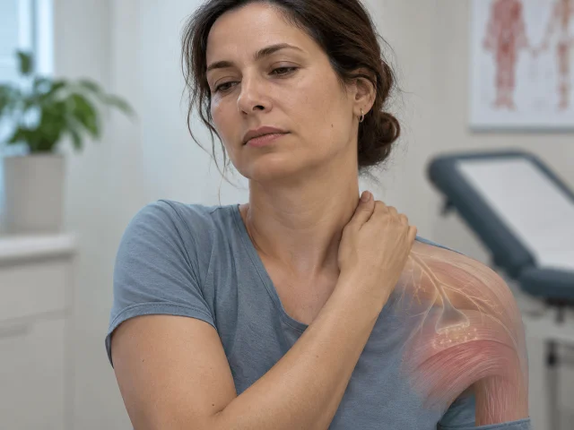
Both shoulders may be affected. Hip symptoms extend to the groin, outer hip or thigh, and the 2023 clinical review also describes neck and upper-arm involvement that makes the distribution broader than pain within an isolated shoulder joint (Florescu et al., 2023). The distribution helps clinicians distinguish widespread inflammatory symptoms from pain confined to an individual injured structure.
This age association is unusually strong. NIAMS reported in 2022 that polymyalgia rheumatica and its related vascular disorder occur almost exclusively beyond age 50, commonly in the late sixties and seventies. The age boundary in classification research is an entry condition, although that research was designed to standardise study groups rather than to confirm diagnoses in individuals (Dasgupta et al., 2012).
Age and population patterns
Raheel and colleagues (2017) identified 377 new cases in Olmsted County, Minnesota, during the study period. In that 2017 population study, 64% of cases were female and mean age at onset was 74.1 years. The cohort was drawn from one Minnesota county.
The same investigators reported an age- and sex-adjusted annual incidence of 63.9 per 100,000 residents aged 50 years and older, with a 95% confidence interval of 57.4–70.4. Incidence increased with age in women and men, although it declined after age 80 in this population (Raheel et al., 2017). The cohort measured incidence rather than biological triggers.
Sex and ancestry also vary across patient groups. NIAMS describes higher occurrence among women and people of Northern European ancestry, while acknowledging cases in other groups (NIAMS, 2022). Specific inherited and environmental causes remain uncertain (NIAMS, 2022).
Polymyalgia rheumatica symptoms and the pattern of morning stiffness
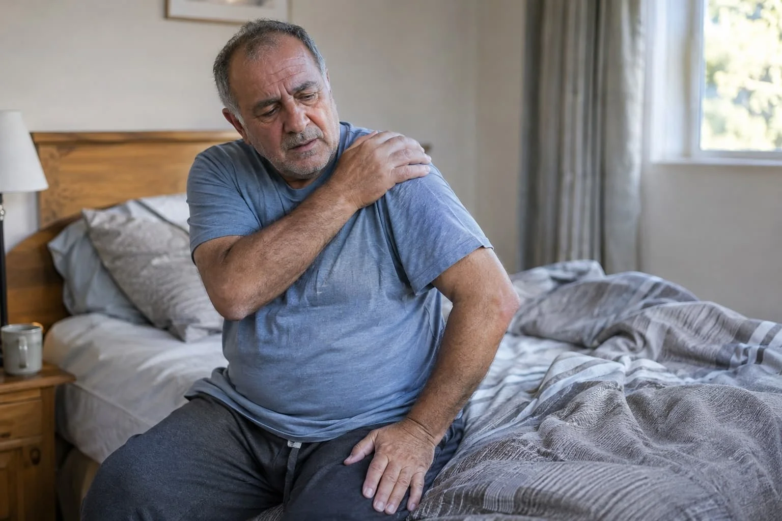
Symptoms often appear quickly. Florescu and colleagues (2023) describe onset over several days, sometimes abruptly overnight. NIAMS (2022) describes stiffness after sleep or inactivity, with symptoms tending to improve during activity.
The painful area may feel like muscle, although the anatomical source can lie around nearby joints and tendons. Several inflammatory conditions present with shoulder discomfort, so symptom location by itself cannot distinguish polymyalgia rheumatica from other causes (Dasgupta et al., 2012). The accompanying duration of stiffness supplies additional information about the pattern.
Shoulder and hip symptoms
Lifting the arms can hurt. Florescu and colleagues (2023) describe a broad shoulder-girdle distribution in which stiffness limits active movement, including the ordinary act of raising the upper arms, while pain does not consistently identify a single injured structure. Difficulty moving is often related to pain rather than demonstrable failure of muscle strength.
Hip symptoms can involve the groin, lateral hip or posterior thigh. This differs from assuming that every symptom described as hip pain arises inside the hip joint (Florescu et al., 2023). Imaging research has found inflammation in periarticular tissues, which gives an anatomical reason for discomfort around a joint without requiring destructive joint disease.
The diagnostic cohort reported by Dasgupta and colleagues (2012) used morning stiffness lasting >45 minutes as an element in its classification algorithm. The threshold belongs to that research instrument and is not an independent proof of disease. Symptoms can still overlap with common shoulder disorders and rheumatoid arthritis.
Functional limitations and systemic symptoms
Ordinary movements can become difficult. In a 2023 review, examples included dressing, brushing hair, rising from a chair and getting out of bed, alongside disrupted sleep due to discomfort (Florescu et al., 2023). Such limitations reflect what the patient experiences during movement rather than a fixed loss of muscle power.
Symptoms extend beyond the joints. NIAMS (2022) lists low-grade fever, reduced appetite and weight loss among accompanying features of polymyalgia rheumatica, conditions that may appear alongside marked morning stiffness and interfere with the interpretation of otherwise non-specific illness symptoms. Fever and weight loss also occur in other illnesses.
Pain can restrict movement. Florescu and colleagues (2023) distinguish discomfort from demonstrable muscle weakness, explaining why the complaint of difficulty raising an arm does not independently establish a primary muscle disorder such as inflammatory myopathy. This distinction matters when considering inflammatory myopathies, which are among the conditions considered during differential diagnosis.
Why polymyalgia rheumatica causes stiffness after age 50
The initiating trigger is unknown. NIAMS (2022) identifies inflammation as the cause of the disorder while stating that researchers have not identified what starts it, leaving genetic, immune and age-related hypotheses to be tested separately. Benlidayi (2024) proposes several ageing-related processes, including immune senescence, as possible contributors.
Inflammation can affect the structures that allow the shoulder and hip girdles to move smoothly. Imaging has located abnormalities around tendons and bursae, while immune studies have reported alterations in circulating and local inflammatory signals (Florescu et al., 2023). No single inflammatory trigger has been established (NIAMS, 2022).
Inflammation around joints and tendons
A bursa is a fluid-containing structure that helps adjacent tissues glide. In polymyalgia rheumatica, ultrasound studies frequently describe subacromial or subdeltoid bursitis and inflammation of the long-head biceps tendon sheath (Florescu et al., 2023). The abnormalities involve bursae and tendon sheaths.
Inflammatory patterns extend beyond the shoulder. Magnetic resonance imaging discussed by Florescu and colleagues (2023) identified affected pelvic tendons and adjacent tissues, offering an anatomical explanation for hip-girdle discomfort that may occur without erosive arthritis. The review also notes that imaging methods differ in access and diagnostic performance.
Immune signalling is involved in polymyalgia rheumatica, but its mechanisms should not be equated with those of the conditions discussed in this overview of autoimmune diseases. Florescu et al. (2023) report a correlation between higher interleukin-6 concentrations and disease activity. That association does not establish whether interleukin-6 initiates the disease or reflects inflammation already taking place.
Ageing of the immune system
Immune ageing has several proposed components. Benlidayi's 2024 narrative review names immunosenescence, age-associated chronic inflammation, endocrine change, infection susceptibility and altered gut microbiota as possible contributors to the concentration of cases in later life. The review raises candidate explanations rather than showing which process starts an individual case.
Investigations of T cells have reported altered distributions in patients with polymyalgia rheumatica or giant cell arteritis. In the 2023 review, an increase in T-helper 17 cells accompanied a decrease in regulatory T cells in a studied group (Florescu et al., 2023). Because the group included both diagnoses, the observation cannot isolate mechanisms specific to polymyalgia rheumatica.
Hormonal ageing offers another hypothesis. Changes in adrenal and other endocrine responses have been proposed as contributors, alongside the inflammatory activity described in affected tissues (Florescu et al., 2023). The endocrine hypothesis has not been demonstrated as an initiating cause.
How polymyalgia rheumatica is diagnosed and distinguished from arthritis
No definitive diagnostic test has been established for polymyalgia rheumatica. Symptoms, inflammatory markers and imaging may support the assessment, but other illnesses can produce overlapping findings (Florescu et al., 2023). A good response to glucocorticoids also cannot be treated as a stand-alone confirmation of the diagnosis (Dasgupta et al., 2012).
Classification rules provide a useful illustration of the limits. Dasgupta and colleagues (2012) designed the criteria for classification in research and explicitly did not test them as individual diagnostic rules. Clinical assessment therefore remains distinct from applying a numerical score.
Blood tests and ultrasound
Erythrocyte sedimentation rate and C-reactive protein are markers of inflammation. The 2012 classification study treated an abnormal result in either marker as part of its entry requirements, together with a characteristic shoulder presentation (Dasgupta et al., 2012). Both markers measure inflammation rather than a unique PMR process.
Dasgupta and colleagues followed 125 patients newly assessed as having polymyalgia rheumatica and 169 comparison patients with mimicking disorders during a six-month prospective cohort (2012). Their clinical classification score reached 68% sensitivity and 78% specificity against all comparison subjects. Specificity was lower when rheumatoid arthritis was the comparison diagnosis, at 65%.
Adding ultrasound criteria changed the balance of classification performance. In the same research, the ultrasound-inclusive algorithm had 66% sensitivity and 81% specificity (Dasgupta et al., 2012). The difference describes performance within the study sample, rather than the predictive value of an isolated scan in routine practice.
Study | Population or design | Measurement | Reported finding |
Raheel et al., 2017 | Minnesota incident PMR cohort | Age and incidence | 377 cases; 64% female |
Dasgupta et al., 2012 | 125 PMR cases and 169 comparators | Classification score | 68% sensitivity; 78% specificity |
Dasgupta et al., 2012 | Ultrasound classification | Sensitivity and specificity | 66% and 81%, respectively |
Spiera et al., 2023 | Relapsing PMR trial | Cumulative glucocorticoid exposure at 52 weeks | 777 mg versus 2044 mg |
Conditions with overlapping symptoms
Rheumatoid arthritis is a major alternative because it can also produce inflammatory pain and stiffness. The 2012 classification study found that distinguishing polymyalgia rheumatica from rheumatoid arthritis was harder than distinguishing it from several local shoulder conditions (Dasgupta et al., 2012). Rheumatoid arthritis was a difficult comparator in that cohort.
The broader differential includes inflammatory myopathies, osteoarthritis, rotator cuff disorders, thyroid disease and infection (Florescu et al., 2023). Osteoarthritis can also involve pain and stiffness, although its clinical features and underlying mechanisms differ from those of polymyalgia rheumatica. A related study digest on osteoarthritis discomfort examines that separate condition rather than providing evidence about polymyalgia rheumatica.
A normal inflammatory blood test can also complicate interpretation. In the Dasgupta et al. (2012) cohort, only five participants lacked abnormal C-reactive protein or erythrocyte sedimentation rate, and the authors described greater diagnostic uncertainty in that setting. Classification performance does not establish absolute exclusion.
How polymyalgia rheumatica is treated and why symptoms can return
Glucocorticoids remain the principal treatment, according to Iorio and colleagues (2025). Iorio and colleagues (2025) associate prolonged glucocorticoid use with toxicity, while Toyoda et al. (2026) describe treatment courses extended by relapse. Spiera and colleagues (2023) also examined recurrence during reduced glucocorticoid exposure.
Treatment research examines remission. Biological drugs directed against interleukin-6 signalling, including tocilizumab and sarilumab, have been studied as possible ways of limiting glucocorticoid exposure in selected patients while maintaining control of inflammatory symptoms (Iorio et al., 2025). Results depend on whether patients have newly diagnosed disease or have relapsed during treatment reduction.
Glucocorticoid response and relapses
Clinical improvement after glucocorticoids is common but variable. In the original classification research, Dasgupta and colleagues (2012) cautioned that even clinically typical cases do not respond uniformly, so treatment response cannot reliably define the diagnosis. Early improvement and sustained control are separate questions.
Relapse is a recurring problem during reductions in glucocorticoid treatment. The 2023 sarilumab trial report states that more than half of patients experience a relapse during tapering (Spiera et al., 2023). The 2026 British Society for Rheumatology guideline also describes relapses as common and links them to longer courses of treatment.
Relapse definitions and study populations vary across the literature. Florescu and colleagues (2023) list other inflammatory or mechanical conditions that may resemble a relapse. Attribution therefore matters when comparing relapse rates across studies.
Treatment research and adverse effects
Spiera and colleagues (2023) studied sarilumab for polymyalgia rheumatica recurring during a glucocorticoid taper. The trial found lower cumulative glucocorticoid exposure in the sarilumab group, with median totals of 777 mg versus 2044 mg at 52 weeks. The trial enrolled patients who had relapsed during a taper.
The investigators also reported a significant advantage in sustained remission during the studied reduction strategy (Spiera et al., 2023). Treatment guidelines and reviews discuss interleukin-6 receptor blockers as additional options in selected circumstances, with professional assessment of the risk and benefit balance (Iorio et al., 2025; Buttgereit et al., 2026).
Conventional disease-modifying agents have less consistent evidence. Iorio and colleagues (2025) characterise the evidence for methotrexate as limited relative to emerging findings for some biological treatments. Glucocorticoid toxicity, treatment complications and long-term outcomes remain relevant, since a reduction in cumulative exposure is only one trial endpoint.
The link between polymyalgia rheumatica and giant cell arteritis
Polymyalgia rheumatica and giant cell arteritis occur in overlapping patient groups. NIAMS describes giant cell arteritis as inflammation of arteries, including vessels around the temples and larger branches extending from the aorta (2022). The two conditions can coexist, but shoulder stiffness does not itself establish arterial inflammation.
The relationship is clinically distinctive because the possible complications differ. Polymyalgia rheumatica principally presents with pain and stiffness, while giant cell arteritis may affect arteries supplying the eyes (NIAMS, 2022).
Shared inflammatory features
NIAMS reported in 2022 that about 10 percent of people with polymyalgia rheumatica also have giant cell arteritis, while about 50 percent of those with giant cell arteritis have polymyalgia rheumatica. These estimates run in different directions because they use different patient groups as the denominator. They cannot be combined into one undifferentiated risk figure.
Shared features include inflammatory activity and concentration of disease in older adults. Immune investigations sometimes enrol patients with both diagnoses, including the T-cell observations described by Florescu and colleagues (2023). The studied immune-cell group included both disorders.
Imaging can sometimes reveal large-vessel involvement alongside a polymyalgia presentation. The 2023 Lancet review notes the growing role of imaging in identifying the inflammatory substrate and detecting coexistent giant cell arteritis (Espígol-Frigolé et al., 2023). The presence of overlapping findings does not mean the disorders have identical consequences.
Symptoms associated with vascular complications
Temple headache is a characteristic symptom. NIAMS (2022) also describes scalp tenderness and pain in the jaw during chewing, complaints linked to the arterial condition rather than to the usual restriction of shoulder and hip movements in isolated polymyalgia rheumatica. Giant cell arteritis involves arteries, as NIAMS describes (2022).
Visual symptoms can be transient. NIAMS (2022) reports episodes of double vision or loss of sight affecting either or both eyes, including disturbances that resolve initially but may precede lasting loss of vision in untreated giant cell arteritis. The possibility of a vision-threatening complication distinguishes the clinical stakes of giant cell arteritis from the usual stiffness pattern of isolated polymyalgia rheumatica.
Inflammatory markers can also become difficult to interpret during interleukin-6 receptor blockade. Toyoda et al. (2026) warn that relapse, infection and new giant cell arteritis can occur without raised markers in people taking these agents. A laboratory result under treatment therefore represents a particular physiological context, not an independent description of vascular status.
What is proven about polymyalgia rheumatica and what is not
Polymyalgia rheumatica has a recognisable pattern of proximal pain, prolonged morning stiffness and onset predominantly in later adulthood. Observational and imaging findings support inflammation around the shoulder and hip girdles, although no single diagnostic test is definitive (Florescu et al., 2023; NIAMS, 2022).
Shoulder and hip stiffness is a consistent clinical pattern, but neither its precise cause nor the diagnosis can be settled by one symptom or laboratory result.
The population evidence clarifies who is affected within defined settings. Raheel and colleagues (2017) observed a higher age-related incidence in an Olmsted County cohort, while the classification cohort of Dasgupta and colleagues (2012) showed imperfect separation from other inflammatory disorders. Both findings limit what can be inferred about an individual patient from age or symptoms alone.
The age mechanism remains unresolved. Immune and endocrine ageing are proposed contributors, while the initiating inflammatory trigger is unknown (Benlidayi, 2024; NIAMS, 2022). Florescu and colleagues (2023) describe T-cell findings from groups containing polymyalgia rheumatica and giant cell arteritis, limiting conclusions about each disease separately.
Treatment trials are more direct evidence for selected interventions. In relapsing disease, Spiera and colleagues (2023) reported lower glucocorticoid exposure with sarilumab, without establishing outcomes for every treatment population. Longitudinal symptom studies collect repeated observations after diagnosis, although repeated questionnaires alone cannot separate disease activity from every alternative cause of discomfort (Hamad et al., 2025).
Classification scores and treatment response remain imperfect diagnostic tools. The original criteria authors warned against using their research score as an individual diagnostic rule and against considering glucocorticoid response a reliable defining feature (Dasgupta et al., 2012). More direct studies of tissue-level inflammation and prospectively defined outcomes would be required to distinguish competing accounts of why disease concentrates in older adults.
Frequently asked questions
Can polymyalgia rheumatica affect people under 50?
It is highly unusual. NIAMS describes polymyalgia rheumatica as occurring almost exclusively after age 50, often in the late sixties or seventies (2022). The age boundary was also required in the principal classification cohort (Dasgupta et al., 2012). The criteria were not tested as diagnostic criteria (Dasgupta et al., 2012).
Does polymyalgia rheumatica cause muscle weakness?
Pain and restricted movement are typical, while objective muscle weakness is not characteristic of the condition (Florescu et al., 2023). A painful shoulder can make an arm difficult to raise without demonstrating primary injury to the muscle fibres. Inflammatory myopathies belong among the alternative diagnoses considered when the presentation differs from the usual pattern.
Is polymyalgia rheumatica the same as rheumatoid arthritis?
No. Both may produce inflammatory stiffness, but their clinical patterns and classifications differ. Dasgupta and colleagues (2012) found that distinguishing polymyalgia rheumatica from rheumatoid arthritis was less specific than separating it from non-inflammatory shoulder problems. Their research also showed that ultrasound findings alone could not cleanly distinguish the two conditions.
Can polymyalgia rheumatica resolve without treatment?
A spontaneous resolution cannot be predicted from the evidence used here. Glucocorticoids are the established principal treatment, and symptoms may recur when treatment is reduced (Iorio et al., 2025; Spiera et al., 2023). No untreated recovery period can be inferred from those treatment studies.
Why can inflammatory blood markers be normal in polymyalgia rheumatica?
Normal markers were uncommon in the 2012 classification cohort, where only five polymyalgia rheumatica patients lacked abnormal C-reactive protein or erythrocyte sedimentation rate (Dasgupta et al., 2012). The authors considered the diagnosis less certain in that setting. Under interleukin-6 receptor blockade, inflammatory markers may also fail to rise during relapse or associated vascular disease (Toyoda et al., 2026).
Sources
- Florescu MM, et al. Polymyalgia rheumatica: An update (Review). Experimental and Therapeutic Medicine. 2023;26(6):543. DOI 10.3892/etm.2023.12242.
- Raheel S, Shbeeb I, Crowson CS, Matteson EL. Epidemiology of Polymyalgia Rheumatica 2000–2014 and Examination of Incidence and Survival Trends Over 45 Years: A Population-Based Study. Arthritis Care & Research. 2017;69(8):1282–1285. DOI 10.1002/acr.23132.
- Dasgupta B, Cimmino MA, Maradit Kremers H, et al. 2012 Provisional Classification Criteria for Polymyalgia Rheumatica: A European League Against Rheumatism/American College of Rheumatology Collaborative Initiative. Arthritis & Rheumatism. 2012;64(4):943–954. DOI 10.1002/art.34356.
- Espígol-Frigolé G, Dejaco C, Mackie SL, et al. Polymyalgia rheumatica. The Lancet. 2023;402(10411):1459–1472. DOI 10.1016/S0140-6736(23)01310-7.
- Spiera RF, Unizony S, Warrington KJ, et al. Sarilumab for Relapse of Polymyalgia Rheumatica during Glucocorticoid Taper. New England Journal of Medicine. 2023;389(14):1263–1272. DOI 10.1056/NEJMoa2303452.
- Toyoda T, Bhide S, Boland C, et al. The 2026 British Society for Rheumatology guideline for the treatment of polymyalgia rheumatica. Rheumatology. 2026;65(10):keag502. DOI 10.1093/rheumatology/keag502.
- Hamad N, Muller S, Hider S, et al. Patterns of pain and stiffness over 5 years in polymyalgia rheumatica: results from the PMR Cohort Study. Rheumatology Advances in Practice. 2025;9(3):rkaf060. DOI 10.1093/rap/rkaf060.
- Iorio L, Bond M, Padoan R, Dejaco C. Advances in the treatment of polymyalgia rheumatica. Rheumatology. 2025;64(Suppl 1):i48–i54. DOI 10.1093/rheumatology/keae539.
- Buttgereit F, Boyadzhieva Z, Reisch M, et al. Recommendations of the German, Austrian and Swiss rheumatology and other medical societies for the management of polymyalgia rheumatica. Rheumatology. 2026;65(2):keaf646. DOI 10.1093/rheumatology/keaf646.
- National Institute of Arthritis and Musculoskeletal and Skin Diseases. Polymyalgia Rheumatica and Giant Cell Arteritis. Last reviewed February 2022. https://www.niams.nih.gov/health-topics/polymyalgia-rheumatica-giant-cell-arteritis
- Benlidayi IC. Why is polymyalgia rheumatica a disease of older adults? Explanations through etiology and pathogenesis: a narrative review. Clinical Rheumatology. 2024;43(3):851–861. DOI 10.1007/s10067-023-06708-3.

Ellen Marsh — author
Ellen writes Health & Medicine explainers for Cabdirect, with a focus on public health, disease prevention and the interpretation of medical research for general readers. Her academic background combines biomedical sciences with postgraduate study in populatio...
This article is for general information and is not medical advice. It was reviewed for accuracy by a qualified clinician; decisions about your health should be made with your own doctor.
Related

Betalain-Rich Red Beet Extract and Osteoarthritis Discomfort
Psoriatic arthritis and why a skin condition affects the joints

What autoimmune disease is and why the immune system attacks the body

How the lymphatic system drains fluid and helps fight infection
How the HPV vaccine works and what studies show about its effect
How Graves’ disease antibodies push the thyroid into overdrive

How the tetanus shot protects and why boosters are needed