
















How the lymphatic system drains fluid and helps fight infection
In broad terms, the lymphatic system drains fluid by collecting the water and protein that leak from tiny blood vessels and returning them to venous blood, and it helps fight infection because that stream also delivers microbes, or cells that have swallowed them, to nearby nodes (Moore and Bertram, 2018; Liao and von der Weid, 2015). One estimate puts the fluid entering lymphatic vessels at around 8 litres a day, with roughly half of it reabsorbed into blood within the nodes (Woodcock and Michel, 2021). The vessels also take up fat from the gut, a third job alongside drainage and defence (Liao and von der Weid, 2015).
The sections below follow the fluid on its way. They describe the parts of the system, explain why fluid gathers in tissues and how lymph gets back into circulation, and then follow an infection into a node. Further on come the vessels discovered in membranes around the brains of mice, the consequences of failed drainage and the open questions.

Evidence last checked 19 Nov 2026 · 14 min
What the lymphatic system is made of
It is a one-way web of vessels linked to lymph nodes, plus the fluid inside it, the cells it carries and several lymphoid organs (Angeli and Lim, 2023; Null et al., 2023). One reference chapter divides the transport route into five parts: capillaries, collecting vessels, nodes, trunks and finally ducts (Ozdowski and Gupta, 2023).
Lymphatic capillaries and collecting vessels
The route begins with blind-ended capillaries, which start as closed stumps about 50 μm across and are wider than neighbouring blood capillaries (Moore and Bertram, 2018; Null et al., 2023). Their lining cells overlap loosely, and when tissue pressure climbs, fine anchoring filaments tug the edges apart so that fluid can pass inside (Angeli and Lim, 2023). Once the vessel has filled, the higher pressure within presses the edges together again and keeps lymph from leaking back (Angeli and Lim, 2023). The gaps measure only about 2–3 μm, yet both fluid and whole cells can slip through (Liao and von der Weid, 2015).
Collecting vessels are mainly built for transport. Their lining cells are joined by tight, zip-like seals, and one-way valves, which the capillaries lack, divide them into short chambers known as lymphangions (Angeli and Lim, 2023; Null et al., 2023; Moore and Bertram, 2018).
These vessels reach almost every organ. One reference chapter places lymphatic tissue in nearly all organs, the eye and even the brain among them (Ozdowski and Gupta, 2023). Another notes that brain and spinal tissue were long believed to have no lymphatic vessels until some turned up in the membranes covering the brain (Null et al., 2023).
Lymph nodes, spleen and other lymphoid tissue
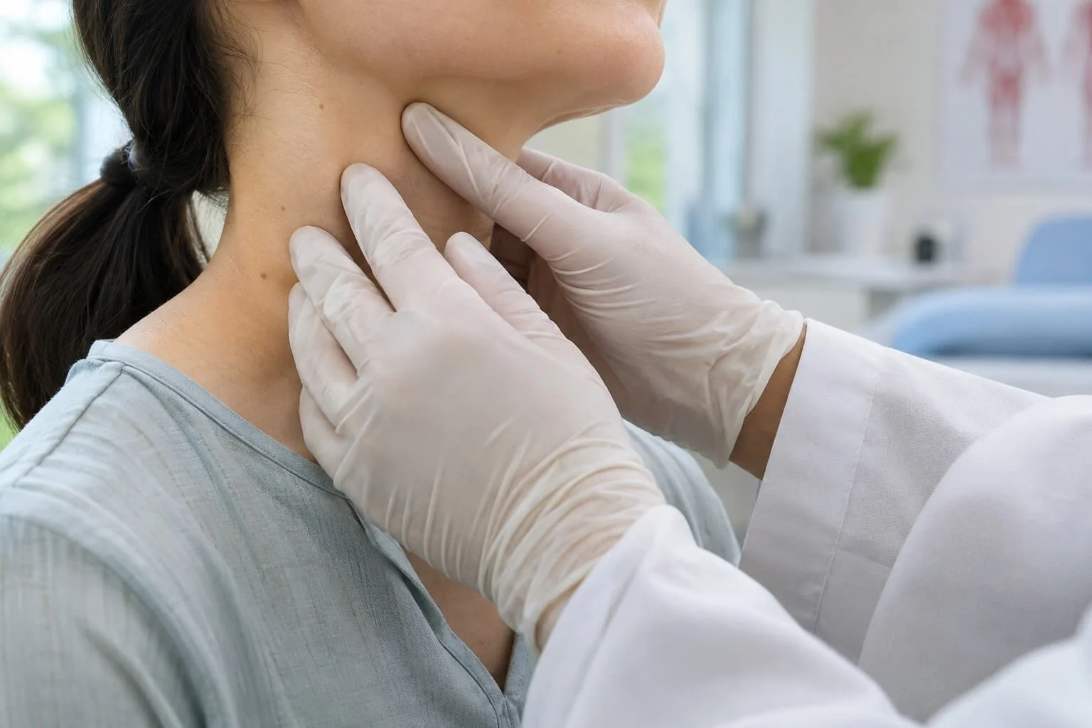
Each lymph node is a small kidney-shaped organ of 1–2 cm or less (Moore and Bertram, 2018). Every drop of lymph crosses at least one node (Moore and Bertram, 2018). Their number is uncertain: a 2018 review gives some 500 to 600, while a reference chapter puts it at about 450 (Moore and Bertram, 2018; Ozdowski and Gupta, 2023). Inside a node, T cells meet dendritic cells, which present antigens, in the paracortex, and B cells gather in follicles in the cortex, the node's outer zone (Ozdowski and Gupta, 2023).
Lymphoid organs are usually split into primary and secondary ones. Lymphocytes are produced in primary organs, meaning the thymus and the marrow inside bones; secondary organs, such as nodes, spleen, tonsils and the gut lining, host the immune work itself (Null et al., 2023). Roughly 80% of all lymph comes from the liver and intestine, according to the same chapter (Null et al., 2023).
Why fluid collects in tissues and becomes lymph
Capillary walls are not watertight. Some plasma water and a share of its proteins pass through them, and this seepage keeps tissues hydrated and supplied with nutrients (Moore and Bertram, 2018).
Filtration from the blood capillaries
For many decades, textbooks described a balance in which fluid left a capillary at its arterial end and was largely drawn back at its venous end, an idea rooted in a hypothesis Ernest Starling put forward in 1896 (Woodcock and Michel, 2021). A 2010 review, combining direct observations with modelling, argued that the evidence does not support lasting reabsorption in most tissues, because a slight outward filtration persists even in venules (Levick and Michel, 2010). The review also treats the glycocalyx, a layer on the inner surface of the vessel wall, as the semipermeable barrier that governs this exchange (Levick and Michel, 2010).
So the lymphatics do most of the return work. In a sentence from the earlier review by Levick and Michel, which Woodcock and Michel later quoted, the fluid balance of most tissues is said to rely heavily on the lymphatics (Woodcock and Michel, 2021). The exceptions named in the modelling review are specialised regions such as the kidney and the intestinal lining, where secretions from nearby epithelium keep absorption going and wash plasma proteins on into the lymphatics (Levick and Michel, 2010).
How much fluid enters the lymphatic system
Estimates of daily volume vary with the source. A 2018 review, drawing on earlier measurements, puts the volume turning into lymph at some 8 litres daily and the flow leaving the nodes at about 4 litres, because water is reabsorbed inside them (Moore and Bertram, 2018). A 2021 review describes the same pattern, with around half of the 8 litres absorbed into the blood within the nodes (Woodcock and Michel, 2021). A StatPearls chapter gives a higher range of 8 to 12 litres a day (Ozdowski and Gupta, 2023).
These figures describe lymph moving through the vessels, not fluid delivered straight to the heart. Lymph leaving a node holds roughly twice as much protein per volume as lymph entering it, a trace of the water removed on the way (Moore and Bertram, 2018).
How lymph travels back to the blood
Pressure in tissue fluid is often lower than atmospheric, whereas the venous junctions that receive lymph sit at about 20 cmH2O, so lymph has to be pushed uphill (Moore and Bertram, 2018). Unlike blood, it has no heart to drive it. Two kinds of pumping do the work instead (Angeli and Lim, 2023; Moore and Bertram, 2018).
Valves and pumping in lymphatic vessels
Intrinsic pumping resembles the action of the heart's chambers (Moore and Bertram, 2018). Each lymphangion contracts its muscular wall while the valves at either end stop backflow, and collecting vessels can repeat these contractions some 10 times a minute; in rat vessel segments studied outside the body the ejection fraction can reach 80% (Moore and Bertram, 2018). In the human leg, contracting vessels with their outflow blocked have produced pressures averaging 61 cmH2O (Moore and Bertram, 2018).
Extrinsic pumping relies on outside pressure, as working muscles, breathing and nearby arterial pulses compress the vessels (Angeli and Lim, 2023; Moore and Bertram, 2018). How the work is shared remains unclear. A reference chapter credits about two-thirds of resting flow to the vessels' own pumping and one-third to muscle compression (Ozdowski and Gupta, 2023). The 2018 review, by contrast, says the share contributed by outside squeezing has never been pinned down and probably varies between organs and activities (Moore and Bertram, 2018).
Where lymph rejoins the bloodstream
Lymph finally drains into one of two large channels, the thoracic duct or the right lymphatic duct (Null et al., 2023). The thoracic duct, the largest lymphatic vessel, is generally thought to carry around three-quarters of what leaves the nodes (Moore and Bertram, 2018). It usually empties low in the neck, at the junction of two veins on the left side, the internal jugular and the subclavian (Null et al., 2023).
Measurements of the duct vary. One chapter gives a diameter of 2 to 6 mm and a length of 36 to 45 cm, whereas the 2018 review reports an average inside diameter of 2.2 mm (Null et al., 2023; Moore and Bertram, 2018).
How lymph nodes help fight infection
Drainage is only half the job. When microbes get into a tissue, the vessels carry the invaders, or immune cells loaded with them, to the closest nodes (Liao and von der Weid, 2015). Riding the flow, this information can arrive in a few tens of minutes, far sooner than cells could crawl there unaided (Moore and Bertram, 2018). For an example of a bacterial infection beginning at the skin barrier, see how staph infections start on the skin.
How antigens and immune cells reach the nodes
Dendritic cells act as messengers. After meeting a microbe they process its antigens, raise their levels of a receptor called CCR7 and speed up towards lymphatic vessels (Liao and von der Weid, 2015). The lymphatic lining makes CCL21, one of the chemical signals CCR7 responds to, and this signal guides the cells in (Liao and von der Weid, 2015; Angeli and Lim, 2023). They pass between the loose junctions of the capillaries and travel with the flow into the outer sinus of the node (Hong et al., 2022).
Classic cannulation studies in people and animals found that T cells account for 80–90% of the cell count in lymph arriving at a node, while dendritic cells and a few B cells make up most of what the review calls the leftover 10–15% (Angeli and Lim, 2023). Size then decides the next step. Small soluble antigens move quickly along narrow conduits deep into the node, while larger particles, bacteria and viruses included, are barred from these channels and swallowed by macrophages along the sinus (Liao and von der Weid, 2015; Card et al., 2014).
Inside the node, dendritic cells present antigens for T cells to inspect and switch on a targeted response (Ozdowski and Gupta, 2023). Many T cells also arrive from the blood through specialised venules and move through the node in an apparently random pattern, which may help them encounter dendritic cells (Hong et al., 2022).
Why lymph nodes swell during infection
A node that is responding to infection has to make room. A 2019 review lists the steps seen mostly in mice: immune cells are drawn in by chemical signals, the supporting network of the node relaxes, its stromal cells divide, the adaptive response unfolds and finally the node contracts (Lucas and Tamburini, 2019). Lymphocytes are also held back for a while, because their exit from an activated node is briefly blocked (Liao and von der Weid, 2015; Card et al., 2014).
The timing in mice does not translate directly to people. In those experiments the lymphatic lining of the node kept expanding between days 4 and 7 after the stimulus, steered by adaptive immunity (Lucas and Tamburini, 2019). How a node shrinks back afterwards has hardly been studied (Lucas and Tamburini, 2019).
Lymphadenitis refers to inflammation of lymph nodes, often associated with a nearby bacterial infection, although viruses and fungi can also be involved (Null et al., 2023). A different type of infection is discussed in the article on why rhinoviruses cause cold symptoms.
Research on lymphatic vessels and immunity
Recent research has changed how scientists regard the system. A 2023 review argues that the network now counts as essential to health rather than as a secondary set of vessels (Angeli and Lim, 2023). A 2014 review made a related point: how lymph vessels move fluid and how they serve immunity, once studied apart, are tightly linked (Card et al., 2014).
Lymphatic vessels in the coverings of the brain
For a long time scientists believed the central nervous system had no conventional route for lymph drainage (Louveau et al., 2015). In 2015 two groups reported lymph vessels running beside the large venous sinuses in the dura mater, the outermost covering of the brain in mice (Louveau et al., 2015; Aspelund et al., 2015). These vessels carried immune cells as well as cerebrospinal fluid and linked up with the deep cervical nodes of the neck (Louveau et al., 2015). The idea was not wholly new: Paolo Mascagni, an Italian anatomist, described such vessels late in the eighteenth century, but no one could repeat his finding (Aspelund et al., 2015).
Human evidence is thinner. In human dura from autopsies, Louveau and colleagues saw only a structure that might be similar and called for further studies (Louveau et al., 2015). In mice that lacked these vessels, the clearance of large molecules from the brain slowed, yet neither fluid pressure in the brain nor its water content changed (Aspelund et al., 2015).
Writing about their mouse work, Aspelund and colleagues noted that the majority of cerebrospinal fluid is still thought to drain into venous blood through arachnoid granulations (Aspelund et al., 2015). The dural vessels found in mice therefore add a route rather than replace one, and both groups called for lymphatics' part in brain physiology and immunity to be reconsidered (Aspelund et al., 2015; Louveau et al., 2015).
Lymphatic endothelium as an active part of immunity
The cells lining lymphatic vessels are probably the first to meet antigens, warning signals and migrating immune cells leaving a tissue (Card et al., 2014). Mouse work shows why this matters. In mice bred without lymphatic capillaries in the skin, vaccination into the skin produced a much weaker antibody response (Card et al., 2014). Blocked lymph flow, the authors concluded, stops antigens from reaching the nodes, which affects both defence and the tolerance of harmless substances (Card et al., 2014).
Inflammation changes the vessels too, though the direction of change varies. Card and colleagues describe how inflammatory signals and faster fluid flow after injury or infection raise lymph formation as well as how efficiently collecting vessels pump (Card et al., 2014). Work in mice, rats and guinea pigs summarised by Liao and von der Weid, by contrast, found pumping strongly suppressed during inflammation (Liao and von der Weid, 2015). A reference chapter suggests that weaker pumping may partly explain the swelling of inflamed tissue (Ozdowski and Gupta, 2023).
Measure | Basis | Figure |
Fluid becoming lymph each day | Estimate from earlier measurements | About 8 L (Moore and Bertram, 2018) |
Lymph leaving the nodes each day | Same estimate | About 4 L (Moore and Bertram, 2018) |
Number of lymph nodes | Review and reference chapter | About 450 to 600 (Moore and Bertram, 2018; Ozdowski and Gupta, 2023) |
Lymphangion contractions | Review estimate | Some 10 a minute (Moore and Bertram, 2018) |
T cells in incoming lymph | Cannulation work in humans and animals | 80–90% (Angeli and Lim, 2023) |
Lymphoedema of the arm after breast cancer | 72 studies pooled | 16.6% (DiSipio et al., 2013) |
People infected with filariasis | Global estimate for 2018 | 51 million (World Health Organization, 2024) |
Note: the figures come from very different kinds of evidence, from laboratory physiology to global health estimates (Moore and Bertram, 2018; World Health Organization, 2024). They describe averages or estimates for groups and say nothing about the lymphatic health of any one person.
When lymphatic drainage fails
When lymph is not carried away, protein-rich fluid builds up in the tissues and leads to swelling, inflammation, growth of fatty tissue and scarring, a condition called lymphoedema (Moore and Bertram, 2018). People with lymphoedema are prone to infections of the soft tissues, because local immune defence depends on lymph flow (Moore and Bertram, 2018). Secondary lymphoedema follows surgery, radiotherapy, infection or injury that damages the vessels, and it is chronic and progressive, with no cure so far (Angeli and Lim, 2023).
Lymphoedema after cancer treatment
Breast cancer treatment is a common cause. A 2013 meta-analysis of studies published from 2000 to 2012 found a pooled arm lymphoedema incidence of 16.6% across 72 studies (DiSipio et al., 2013). Limited to 30 prospective cohort studies, the estimate rose to 21.4%, which the authors read as over one in five breast cancer survivors (DiSipio et al., 2013). Incidence seemed to climb during the first 2 years after surgery or diagnosis (DiSipio et al., 2013).
The type of surgery made a clear difference. Lymphoedema was about four times as common once the armpit lymph nodes were removed, at 19.9%, as after sentinel-node biopsy (DiSipio et al., 2013). Extensive surgery and excess body weight had the strongest evidence as risk factors (DiSipio et al., 2013).
Lymphatic filariasis
Worldwide, a parasitic infection is another major cause of damaged drainage. Lymphatic filariasis, also called elephantiasis, is spread by mosquitoes, and the adult worms lodge in lymph vessels and impair their function (World Health Organization, 2024). The World Health Organization counted 51 million infections as of 2018, a fall of 74% since its elimination programme began in 2000 (World Health Organization, 2024). Some 657 million people across 39 countries lived in 2023 in areas where whole communities need preventive treatment (World Health Organization, 2024).
At least 36 million individuals still live with chronic effects such as lymphoedema or hydrocele (World Health Organization, 2024). The acute episodes are mostly caused by bacterial skin infections that set in where lymphatic damage has weakened local defences (World Health Organization, 2024).
What is established about the lymphatic system and what remains uncertain
Several points are well established. Lymphatic vessels carry back fluid that has escaped from capillaries, take up fat from food and deliver antigens, and the cells that carry them, to the nodes (Liao and von der Weid, 2015; Ozdowski and Gupta, 2023). Physiological reviews and modelling suggest that in most tissues the lymphatics, not reabsorption into capillaries, are the main route back for filtered fluid (Levick and Michel, 2010; Woodcock and Michel, 2021).
Lymph can deliver signals to the nodes in a matter of tens of minutes, and in mice lacking lymphatic capillaries in the skin a skin vaccine produced a much weaker antibody response (Moore and Bertram, 2018; Card et al., 2014).
Other questions remain open. Basic figures differ between sources, from the number of lymph nodes to the daily volume of lymph (Moore and Bertram, 2018; Ozdowski and Gupta, 2023). How lymphatic pumping and flow shape immune responses in cancer and in long-term inflammation remains unanswered (Liao and von der Weid, 2015). A review of mostly mouse studies adds that how lymphatic cells balance immune tolerance against protective immunity also remains poorly understood (Lucas and Tamburini, 2019).
The papers cited here, including the mouse studies, acknowledge a mix of public and charitable funding, from the US National Institutes of Health and the European Research Council to private foundations (Liao and von der Weid, 2015; Card et al., 2014; Louveau et al., 2015). The 2013 meta-analysis was funded by the National Breast Cancer Foundation in Australia (DiSipio et al., 2013).
Frequently asked questions
What is lymph made of?
Lymph begins as the fluid around the cells, so it is watery, clear and yellowish, much like blood plasma (Null et al., 2023). Before reaching a node it carries water, salts, plasma proteins and white cells (Moore and Bertram, 2018). Gut lymph, called chyle, looks milky because of fat droplets called chylomicrons (Moore and Bertram, 2018).
Is the lymphatic system part of the immune system?
It belongs to both systems. A StatPearls chapter places it within the circulation while giving it a critical role in immune function and fluid drainage (Null et al., 2023). Its nodes, spleen and tonsils are secondary lymphoid organs where immune cells do their work (Null et al., 2023).
Do swollen lymph nodes always mean an infection?
No. Nodes can change in size or texture in inflammatory, infectious, autoimmune or malignant conditions, known as lymphadenopathy (Ozdowski and Gupta, 2023). Inflamed nodes usually follow a nearby bacterial infection, though viruses and fungi can be involved (Null et al., 2023). Swelling alone does not show which condition is responsible (Ozdowski and Gupta, 2023).
How many lymph nodes does the human body have?
Estimates differ. A 2018 review gives some 500 to 600, while a StatPearls chapter puts the number at about 450 (Moore and Bertram, 2018; Ozdowski and Gupta, 2023). They are small kidney-shaped organs of 1–2 cm or less, and all lymph passes through at least one (Moore and Bertram, 2018).
Does the lymphatic system have its own pump?
Not a central one. Muscle cells in the walls of collecting vessels contract rhythmically, and one-way valves stop backflow (Angeli and Lim, 2023; Null et al., 2023). Nearby skeletal muscles and pulsing arteries add outside squeezing (Angeli and Lim, 2023). The relative share of each mechanism is uncertain (Moore and Bertram, 2018).
Sources
- Liao S, von der Weid PY. Lymphatic system: an active pathway for immune protection. Seminars in Cell & Developmental Biology. 2015;38:83–89. DOI 10.1016/j.semcdb.2014.11.012.
- Angeli V, Lim HY. Biomechanical control of lymphatic vessel physiology and functions. Cellular & Molecular Immunology. 2023;20(9):1051–1062. DOI 10.1038/s41423-023-01042-9.
- Null M, Arbor TC, Agarwal M. Anatomy, Lymphatic System. In: StatPearls. Treasure Island (FL): StatPearls Publishing; 2023 (updated 6 March 2023). NCBI Bookshelf NBK513247.
- Moore JE Jr, Bertram CD. Lymphatic system flows. Annual Review of Fluid Mechanics. 2018;50:459–482. DOI 10.1146/annurev-fluid-122316-045259.
- Levick JR, Michel CC. Microvascular fluid exchange and the revised Starling principle. Cardiovascular Research. 2010;87(2):198–210. DOI 10.1093/cvr/cvq062.
- Woodcock TE, Michel CC. Advances in the Starling principle and microvascular fluid exchange; consequences and implications for fluid therapy. Frontiers in Veterinary Science. 2021;8:623671. DOI 10.3389/fvets.2021.623671.
- Hong W, Yang B, He Q, Wang J, Weng Q. New insights of CCR7 signaling in dendritic cell migration and inflammatory diseases. Frontiers in Pharmacology. 2022;13:841687. DOI 10.3389/fphar.2022.841687.
- Card CM, Yu SS, Swartz MA. Emerging roles of lymphatic endothelium in regulating adaptive immunity. Journal of Clinical Investigation. 2014;124(3):943–952. DOI 10.1172/JCI73316.
- Lucas ED, Tamburini BAJ. Lymph node lymphatic endothelial cell expansion and contraction and the programming of the immune response. Frontiers in Immunology. 2019;10:36. DOI 10.3389/fimmu.2019.00036.
- Louveau A, Smirnov I, Keyes TJ, Eccles JD, Rouhani SJ, Peske JD, Derecki NC, Castle D, Mandell JW, Lee KS, Harris TH, Kipnis J. Structural and functional features of central nervous system lymphatic vessels. Nature. 2015;523(7560):337–341. DOI 10.1038/nature14432.
- Aspelund A, Antila S, Proulx ST, Karlsen TV, Karaman S, Detmar M, Wiig H, Alitalo K. A dural lymphatic vascular system that drains brain interstitial fluid and macromolecules. Journal of Experimental Medicine. 2015;212(7):991–999. DOI 10.1084/jem.20142290.
- DiSipio T, Rye S, Newman B, Hayes S. Incidence of unilateral arm lymphoedema after breast cancer: a systematic review and meta-analysis. Lancet Oncology. 2013;14(6):500–515. DOI 10.1016/S1470-2045(13)70076-7.
- World Health Organization. Lymphatic filariasis. Fact sheet, 21 November 2024. Geneva: WHO. https://www.who.int/news-room/fact-sheets/detail/lymphatic-filariasis
- Ozdowski L, Gupta V. Physiology, Lymphatic System. In: StatPearls. Treasure Island (FL): StatPearls Publishing; 2023 (updated 1 May 2023). NCBI Bookshelf NBK557833.

Nora Veldin — author
Nora Veldin is a health science writer with academic training in biomedical science and public health. After completing a BSc in Biomedical Science, she undertook an MPH focused on epidemiology, population health and the interpretation of health evidence. Her...
This article is for general information and is not medical advice. It was reviewed for accuracy by a qualified clinician; decisions about your health should be made with your own doctor.
Related
What autoimmune disease is and why the immune system attacks the body
How staph infections start on the skin and reach the blood
Common cold symptoms and why rhinoviruses cause them

Polymyalgia rheumatica and why shoulders and hips stiffen after 50
How the HPV vaccine works and what studies show about its effect
How Graves’ disease antibodies push the thyroid into overdrive
How the tetanus shot protects and why boosters are needed