





Chest disease diagnosis: radiologic signs and differentials
Chest disease is diagnosed by combining the clinical story with targeted tests rather than by treating any single image or laboratory result as decisive. Chest radiography remains a common first imaging test for many respiratory presentations, while CT, ultrasound, vascular imaging, pulmonary function testing and tissue sampling answer specific diagnostic questions. Radiologic signs such as consolidation, ground-glass opacity, atelectasis, hyperlucency, nodules, pleural fluid or mediastinal widening narrow the possibilities, but most are patterns shared by several diseases.
The 1999 fourth edition of *Diagnosis of Diseases of the Chest* reflects a diagnostic framework built around clinical assessment, radiologic signs, physiologic testing and differential diagnosis. Modern practice follows the same core principles while incorporating advances in CT imaging, lung-function assessment, vascular imaging and minimally invasive tissue sampling. Clinicians now combine pattern, distribution, time course and clinical context to select appropriate tests and distinguish between competing causes of chest disease. These principles provide a practical framework for understanding common thoracic imaging findings and the diagnostic pathways used in current respiratory medicine.
Evidence last checked 25 Sep 2026 · 12 min
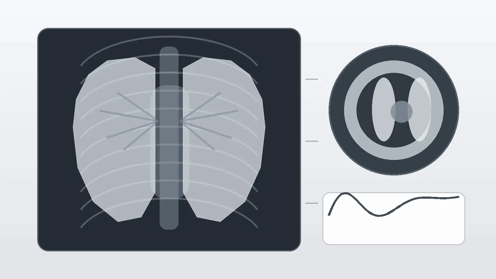
Diagnosis starts with the clinical question
The same radiographic pattern can have very different meanings in two patients, so the diagnostic process begins before an image is acquired. Symptoms such as cough, dyspnoea, fever, haemoptysis and pleuritic pain are interpreted alongside their onset, duration and severity. Age, smoking history, occupational or environmental exposure, immune status, recent surgery, travel, medications and previous cancer can materially change the pre-test probability of infection, malignancy, vascular disease or chronic airway disease. Physical examination can add clues such as unilateral breath-sound reduction, crackles, wheeze, signs of consolidation or evidence of fluid overload, but it rarely identifies a thoracic diagnosis with enough certainty on its own. Clinical history, examination, imaging, physiology and pathology work as complementary parts of the diagnostic process rather than competing alternatives.
A useful diagnostic question is therefore specific: is there focal infection, diffuse parenchymal disease, airway obstruction, pleural disease, a vascular event, a mass, or another explanation outside the lungs? The test is chosen to answer that question with the least unnecessary risk. This is why a chest X-ray can be appropriate as an initial study for many patients with acute respiratory illness or chronic cough, while CT becomes more useful when radiography is negative or indeterminate despite persistent clinical concern, or when diffuse lung disease is suspected. A suspected pulmonary embolism follows a different pathway in which clinical probability and D-dimer testing help determine whether CT pulmonary angiography is appropriate.
Choosing the diagnostic method
Chest radiography and computed tomography
Chest radiography is fast, widely available and useful for surveying the lungs, pleura, heart size, mediastinal contours, diaphragm and visible bones. It can show air-space opacity, lobar collapse, pleural effusion, pneumothorax, large masses and many patterns of chronic lung disease, but a two-dimensional projection can hide lesions behind the heart, diaphragm or other structures. Technique also matters: rotation, low lung volume, underpenetration and portable anteroposterior acquisition can mimic or exaggerate abnormal findings. For this reason, an X-ray is best treated as a structured overview rather than a definitive map of thoracic anatomy.
CT removes much of the superimposition inherent in radiography and characterises attenuation, margins, distribution and anatomy in cross-section. Thin-section CT is central to assessing suspected diffuse lung disease because it can distinguish patterns such as ground-glass opacity, reticulation, honeycombing, emphysema and bronchiectasis far better than a plain film. Contrast-enhanced CT is used when vessels, mediastinal structures, pleura or suspected tumour staging need evaluation; CT pulmonary angiography is tailored to pulmonary arteries. The choice between non-contrast, contrast-enhanced, high-resolution and angiographic protocols is therefore part of the diagnostic reasoning rather than a minor technical detail.
Ultrasound, MRI and nuclear imaging
Thoracic ultrasound is particularly useful when the abnormality reaches the pleural surface. It can confirm and characterise pleural fluid, identify septations, guide drainage or biopsy, and help assess pleural or peripheral lesions in experienced hands. Ultrasound is an important component of evaluating pleural effusion and pleural malignancy, while CT provides a broader view of the entire thorax. Ultrasound is primarily suited to pleural and peripheral abnormalities because aerated lung limits penetration of the sound beam.
MRI has no ionising radiation and offers excellent soft-tissue contrast, although lung imaging is technically challenging because of respiratory motion and the low proton density of aerated lung. It is valuable in selected questions involving the mediastinum, chest wall, diaphragm, heart or vascular structures, and increasingly in specialised lung applications. Ventilation-perfusion scintigraphy can be used when pulmonary embolism is suspected and CT pulmonary angiography is not suitable for the patient. Nuclear medicine and PET-CT can also provide functional or metabolic information when the clinical question extends beyond anatomy, particularly in oncologic staging.
Pulmonary function, bronchoscopy and tissue diagnosis
Imaging describes structure; pulmonary function tests describe physiology. Spirometry measures airflow and forced volumes and is fundamental in assessing obstructive disorders, while lung volumes and gas-transfer testing can clarify restriction, hyperinflation or impaired diffusion when indicated. Modern respiratory testing standards emphasise test quality, reference limits and interpretation in clinical context rather than relying on a single percentage-of-predicted threshold. An abnormal physiologic pattern can support the diagnosis and quantify impairment, while the underlying cause is determined by combining functional results with clinical and structural findings.
Bronchoscopy allows direct airway inspection and sampling by washing, brushing or biopsy. Endobronchial ultrasound can sample lymph nodes and lesions adjacent to major airways, while CT-guided percutaneous biopsy is often suited to peripheral lesions. In suspected lung cancer, CT information helps guide biopsy planning and selection of a sampling route that can provide diagnosis and staging with the least risk. Histology, cytology and microbiology can then define the nature of lesions identified through clinical assessment and imaging.
How radiologic signs narrow the differential diagnosis
Radiologic terminology is most useful when it describes what is visible without prematurely naming the cause. Standardised thoracic imaging terminology allows words such as opacity, nodule, cavity, ground-glass opacity and honeycombing to convey reproducible imaging patterns. A sign becomes diagnostically valuable when it is combined with its location, symmetry, morphology, associated findings and clinical setting. A lower-lobe opacity with fever, for example, is approached differently from the same opacity in a patient with known malignancy or recent aspiration.
Increased density and air-space patterns
Increased lung density on radiography or CT may reflect replacement of air, collapse, fluid, cells or tissue. Consolidation is an air-space opacity that obscures underlying vessel margins more completely than ground-glass opacity; an air bronchogram may be visible when air-filled bronchi remain within opaque lung. Pneumonia is one possible cause, but pulmonary oedema, haemorrhage, aspiration and some tumours can produce overlapping appearances. Ground-glass opacity on CT means increased attenuation in which bronchial and vascular margins remain visible. It is a descriptive imaging term and can occur in infection, oedema, inflammation, partial alveolar filling, early fibrosis and other processes.
Volume loss, decreased density and abnormal air
Atelectasis describes reduced lung volume and is recognised by displacement: fissures may move, the hilum or mediastinum may shift toward the affected side, and adjacent structures can become crowded. The differential includes airway obstruction by mucus or tumour, compression from pleural fluid or mass effect, and postoperative or shallow-breathing states. By contrast, hyperlucency or reduced attenuation can result from emphysema, air trapping, reduced perfusion, pneumothorax or technical factors. A true pneumothorax is suggested by a visceral pleural line with absent peripheral lung markings, while skin folds, bedding and other artefacts can imitate that appearance on portable films.
Nodules, masses, cavities and interstitial patterns
A pulmonary nodule is a focal rounded opacity no larger than 3 cm; larger focal lesions are termed masses in standard thoracic imaging terminology. Size alone does not identify benignity or malignancy. Margin, density, calcification, growth on prior imaging and patient risk factors all influence the differential and help determine whether follow-up, PET-CT or tissue sampling is appropriate. Cavitation describes a gas-filled space within a consolidation, mass or nodule and can occur with infection, malignancy, inflammatory disease and infarction. Reticulation, septal thickening and honeycombing point toward interstitial processes and are interpreted according to their distribution and associated findings because conditions such as oedema and fibrosis can both create line-based patterns.
Pleural and mediastinal signs
Pleural fluid can blunt a costophrenic angle or form a meniscus on upright radiography, while ultrasound is more sensitive for small fluid collections and helps distinguish free-flowing from complex fluid. Pleural thickening, nodularity or unilateral effusion may raise concern for malignancy but are not specific. CT, ultrasound, fluid analysis and tissue sampling can be combined according to the clinical setting to characterise pleural abnormalities. Mediastinal widening is similarly nonspecific: rotation, magnification on anteroposterior films, vascular enlargement, lymphadenopathy, mass and haemorrhage can all alter the contour. Cross-sectional imaging is often used when a mediastinal abnormality is considered clinically important.
Common imaging patterns and what they can mean
Pattern or sign | What it describes | Common diagnostic categories | What helps next |
Consolidation | Dense air-space opacity, sometimes with air bronchograms | Infection, oedema, haemorrhage, aspiration, tumour | Distribution, symptoms, CT, microbiology |
Ground-glass opacity | Increased CT attenuation with vessels still visible | Infection, oedema, inflammation, haemorrhage, early fibrosis | Pattern, chronicity, associated CT signs |
Atelectasis | Loss of lung volume with displacement of structures | Airway obstruction, compression, postoperative change | Assessment for an obstructing lesion or pleural process |
Hyperlucency | Reduced density or increased air | Emphysema, air trapping, pneumothorax, reduced perfusion | Technique assessment, expiratory imaging, vascular signs |
Nodule or mass | Focal rounded pulmonary opacity | Benign lesion, primary cancer, metastasis, infection | Prior imaging, CT morphology, risk assessment, PET-CT or biopsy |
Cavity | Gas-filled space within a nodule, mass or consolidation | Infection, malignancy, inflammatory disease, infarction | Wall features, distribution, microbiology, tissue analysis |
Pleural effusion | Fluid in the pleural space | Heart failure, infection, malignancy, embolic disease and others | Ultrasound, fluid analysis, CT when indicated |
Mediastinal widening | Broadened mediastinal contour | Technique, vascular enlargement, lymph nodes, mass, haemorrhage | Projection assessment and cross-sectional imaging |
Building the differential: five filters that change meaning
A practical differential diagnosis is built by progressively filtering possibilities rather than by memorising a long list for each sign. The most useful filters are the time course, anatomical distribution, patient context, accompanying imaging features and non-imaging data. Acute bilateral ground-glass opacity in a febrile, immunocompromised patient is a different problem from slowly progressive subpleural reticulation in an older adult with exertional dyspnoea. A cavitary upper-lobe lesion in a patient with tuberculosis exposure, a smoker with weight loss, and a patient with septic emboli share a visual feature but not the same prior probabilities. The diagnostic value comes from combining the common sign with the individual clinical context.
- Time course: hours to days favours a different set of causes from changes evolving over months or years.
- Distribution: focal versus diffuse, central versus peripheral, upper versus lower lobe and unilateral versus bilateral patterns matter.
- Associated signs: lymph nodes, pleural fluid, volume loss, airway changes, vascular abnormalities and bone findings can redirect the differential.
- Clinical probability: exposures, immune status, smoking, recent procedures, cancer history and thromboembolic risk alter how an image should be weighted.
- Comparison and confirmation: prior imaging, physiology, laboratory tests, microbiology and tissue sampling can convert a pattern-based differential into a diagnosis.
This approach also reduces common reasoning errors. A single positive imaging sign should not be allowed to override contradictory history or physiology, and a common disease should not be excluded simply because one textbook feature is absent. Conversely, an unusual diagnosis should not be preferred just because an image has an eye-catching sign. Good differential diagnosis is probabilistic: it ranks explanations, identifies dangerous alternatives that require prompt consideration and selects the next test most likely to influence management.
Why a normal chest radiograph does not end the investigation
A normal or near-normal chest radiograph can coexist with clinically important disease. Pulmonary embolism is a classic example: radiography may be normal or show nonspecific findings, so diagnosis depends on clinical probability, D-dimer testing when appropriate and vascular imaging rather than on the plain film. Early or subtle diffuse lung disease can also be difficult to identify on radiography because CT is more sensitive to fine interstitial and ground-glass abnormalities. Small pulmonary nodules, early infection, mild bronchiectasis and predominantly airway disorders may also produce subtle or inconspicuous radiographic changes. Persistent symptoms or high-risk clinical features can therefore justify additional testing despite an apparently reassuring X-ray.
The opposite problem also occurs: an abnormal film does not automatically prove active disease. Old scarring, healed granulomas, benign calcification, technical artefact and normal anatomical variants can create apparent abnormalities. Comparison with previous studies is often one of the highest-value steps because stability over time can sharply alter the differential. When an abnormality is new, progressive or unexplained, the next investigation is selected according to the clinical question and the type of abnormality identified.
What has changed since the 1999 reference text
Diagnostic approaches described in the late 1990s already integrated normal anatomy, investigative methods, radiologic signs and disease-based differential diagnosis. Modern practice has since shifted in important ways. Multidetector CT now provides faster and higher-resolution cross-sectional imaging; CT pulmonary angiography has become central to pulmonary embolism work-up; PET-CT has a larger role in cancer staging; endobronchial ultrasound offers minimally invasive lymph-node sampling; and thin-section CT terminology has become increasingly standardised. Pulmonary function interpretation has also moved toward updated reference equations, z-scores and explicit quality criteria.
These developments preserve the underlying diagnostic logic while improving the precision with which individual clinical questions can be investigated. The durable principle is that morphology and physiology should be linked to clinical probability. Modern diagnostic standards make that relationship more explicit by defining situations in which particular imaging methods are appropriate, incorporating pre-test probability into vascular diagnosis and using biopsy strategies designed to obtain useful diagnostic and staging information with the least risk. Contemporary practice therefore combines established diagnostic principles with current imaging, physiologic testing and tissue-sampling techniques.
What the combined evidence supports
Modern chest diagnosis is based on a method rather than a single universally preferred test. Chest radiography is a valuable first survey in many common presentations, while CT is the main problem-solving tool when anatomical detail, distribution or staging matters. Specialised CT protocols are selected for vascular, interstitial or contrast-enhanced questions. Ultrasound is particularly useful for pleural disease and procedures, pulmonary function testing defines physiologic impairment, and bronchoscopy or percutaneous biopsy provides tissue for histologic, cytologic or microbiological assessment. Each technique contributes a different part of the diagnostic picture.
Radiologic signs should therefore be read as structured observations: what changed, where it changed, how extensive it is and which accompanying features are present. Differential diagnosis then combines those observations with the patient's time course, symptoms and risk profile before determining the most appropriate next investigation. That framework helps explain why two patients with similar X-rays may require different diagnostic pathways, and why a normal X-ray can still lead to CT, vascular imaging or functional testing when clinically indicated.
The goal is not simply to label an image; it is to reach the most defensible diagnosis through a structured and proportionate diagnostic process.
Frequently asked questions
What is usually the first imaging test for chest symptoms?
Chest radiography is a common first imaging test for many respiratory presentations because it is quick and can reveal pneumonia, pleural fluid, pneumothorax, collapse or a large mass. It is not universal, however. The first test depends on the clinical question, urgency and pre-test probability, and some pathways move directly to specialised imaging.
When is CT more useful than a chest X-ray?
CT is more useful when anatomy must be seen in cross-section, when an X-ray is negative or indeterminate despite ongoing concern, or when diffuse lung disease, a mass, mediastinal disease or complications need characterisation. Protocol matters: non-contrast, contrast-enhanced, thin-section and pulmonary angiographic CT answer different diagnostic questions.
Can serious chest disease be present with a normal X-ray?
Yes. Pulmonary embolism may have a normal or nonspecific radiograph, and early interstitial disease, small nodules, mild bronchiectasis or predominantly airway disease can be difficult to see on plain films. A normal X-ray therefore lowers concern for some conditions but does not overrule a strong clinical indication for further testing.
What does ground-glass opacity mean on CT?
Ground-glass opacity is a descriptive CT term for increased lung attenuation through which bronchial and vascular margins remain visible. It is not a diagnosis. Infection, oedema, inflammation, haemorrhage, partial alveolar filling and early fibrosis can all produce it, so distribution, time course and associated findings are needed to narrow the differential.
Why is biopsy sometimes needed after imaging?
Imaging can strongly suggest a category of disease but may not distinguish cancer, infection and inflammatory conditions reliably enough for treatment decisions. Tissue sampling by bronchoscopy, endobronchial ultrasound, image-guided biopsy or another route can provide histology, cytology or microbiology. The safest method depends on lesion location and the information needed for diagnosis and staging.
Sources
- Fraser RS, Müller NL, Colman NC, Paré PD. Fraser and Paré's Diagnosis of Diseases of the Chest. 4th ed. W.B. Saunders; 1999.
- Bankier AA, MacMahon H, Colby T, et al. Fleischner Society: Glossary of Terms for Thoracic Imaging. Radiology. 2024;310(2):e232558. doi:10.1148/radiol.232558.
- American College of Radiology. ACR Appropriateness Criteria: Acute Respiratory Illness in Immunocompetent Patients. Revised 2024.
- American College of Radiology. ACR Appropriateness Criteria: Diffuse Lung Disease.
- Kirsch J, Brown RKJ, Henry TS, et al. ACR Appropriateness Criteria Suspected Pulmonary Embolism: 2022 Update. J Am Coll Radiol. 2022;19(11 Suppl):S488-S501. doi:10.1016/j.jacr.2022.09.014.
- Roberts ME, Rahman NM, Maskell NA, et al. British Thoracic Society Guideline for pleural disease. Thorax. 2023;78(Suppl 3):s1-s42. doi:10.1136/thorax-2022-219784.
- National Institute for Health and Care Excellence. Lung cancer: diagnosis and management. NICE guideline NG122. Updated 8 March 2024.
- Graham BL, Steenbruggen I, Miller MR, et al. Standardization of Spirometry 2019 Update: An Official ATS/ERS Technical Statement. Am J Respir Crit Care Med. 2019;200(8):e70-e88. doi:10.1164/rccm.201908-1590ST.
- Stanojevic S, Kaminsky DA, Miller MR, et al. ERS/ATS technical standard on interpretive strategies for routine lung function tests. Eur Respir J. 2022;60(1):2101499. doi:10.1183/13993003.01499-2021.
- American College of Radiology. ACR Appropriateness Criteria: Chronic Cough.

Tomas Verren — author
Tomas Verren has a background in biomedical sciences and population health, with academic training in human biology, epidemiology, study design and the interpretation of health data. After completing his postgraduate training, he moved into research communicat...
This article is for general information and is not medical advice. It was reviewed for accuracy by a qualified clinician; decisions about your health should be made with your own doctor.
Related

Selenium and cancer prevention: what the evidence shows

Human blood groups and population genetics in Mourant’s atlas