







Equine Anatomy and Clinical Examination in the 2010 Third Edition
The third edition of Praxisorientierte Anatomie und Propädeutik des Pferdes is a 2010 German-language reference devoted to horse anatomy and clinical examination procedures. Written and edited by H. Wissdorf, H. Gerhards, B. Huskamp and E. Deegen, the 1,055-page book is designed to connect detailed anatomical knowledge with the practical requirements of equine veterinary work.
The book covers clinically important areas including the musculoskeletal system and lameness diagnosis, reproduction, the digestive system and examination of the male and female reproductive organs. The edition also adds more than 200 new illustrations, expanded reproductive material, new content on teeth and supporting structures, gastroscopic findings in stomach ulcer and laparoscopic images of abdominal organs.
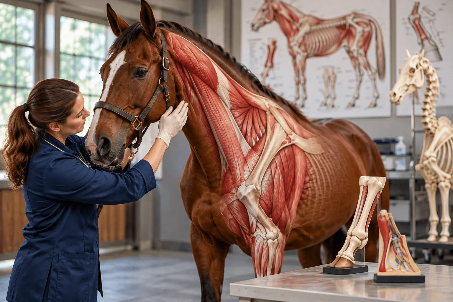
How the Book Connects Equine Anatomy With Clinical Practice
The third edition approaches anatomy as knowledge used during clinical examination rather than as a collection of isolated names and structures. Its substantial length reflects the range of systems and procedures that equine veterinarians encounter. The relationship between an anatomical landmark and the examination performed around it is a recurring organizing principle.
For a student, an anatomical description is most useful when it clarifies where a structure lies, what can be observed or palpated, and why that structure matters during examination. For a practitioner, the same foundation helps communicate findings precisely and organize the next stage of investigation. The book brings these educational and clinical purposes together.
Anatomical knowledge does not replace clinical judgment or a complete diagnostic work-up. Instead, it supplies the spatial and structural framework needed to interpret observations. The third edition can therefore be read as a reference linking basic description with practical examination without presenting any one anatomical finding as a diagnosis.
Table 1. Anatomical Areas and Their Examination Context
Area | Clinical context |
Horse anatomy | Structural foundation for examination |
Musculoskeletal system | Assessment of locomotor abnormalities |
Reproduction | Examination of male and female organs |
Digestive system | Anatomy and gastroscopic observations |
Why Musculoskeletal Anatomy Matters in Equine Lameness
The musculoskeletal system and lameness diagnosis receive particular attention because movement depends on the coordinated function of bones, joints, muscles, tendons and related tissues. Understanding the position and relationships of these structures is a necessary starting point for interpreting a clinical examination. A description of anatomy alone, however, cannot identify the cause of an individual horse’s lameness.
Examination of a lame horse requires the clinician to connect the observed problem with possible anatomical sources. The book’s combined treatment of structure and examination helps explain why knowledge of a region matters when investigating locomotor abnormalities. This is an educational connection rather than a claim that a reference text can replace diagnostic testing.
Anatomical terminology also supports clear communication between clinicians. When findings are described using consistent names for regions and structures, records can be interpreted more reliably. That practical value is one reason a broad anatomical reference remains relevant even as diagnostic methods evolve.
How the Third Edition Expands Reproductive Anatomy
The third edition expands coverage of examination of male and female reproductive organs. It also includes fertilization, placentation, birth, breeding and problems associated with the birth process. Together, these subjects connect the anatomy of reproductive structures with the physiological events and clinical situations in which those structures are examined.
The anatomy of reproductive organs is not interchangeable with the processes of reproduction. Structural knowledge helps explain the location and relationships of organs, while fertilization and placentation concern biological events. Bringing these topics into the same reference allows readers to consider anatomy alongside the practical questions encountered in breeding and reproductive assessment.
Birth-related complications add another clinical dimension. The source highlights their inclusion but does not establish a particular treatment protocol or recommendation. Readers should distinguish the book’s stated scope from contemporary decisions about reproductive intervention, which require current veterinary guidance and patient-specific assessment.
Table 2. Reproductive Topics Highlighted in the Third Edition
Topic | Coverage |
Male and female organs | Expanded examination material |
Fertilization | Included in reproductive discussion |
Placentation | Included in reproductive discussion |
Birth and breeding | Expanded coverage |
Birth-related problems | Clinical complications discussed |
What the Edition Adds on Equine Teeth and Digestion
New material on teeth and their supporting structures extends the reference beyond the regions traditionally emphasized in a general anatomy survey. Dental structures have distinctive relationships to surrounding tissues, and understanding these relationships can assist the organization of an oral examination. The addition indicates greater attention to equine dental anatomy in the third edition.
The digestive system is another clinically relevant subject. The third edition includes gastroscopic findings associated with stomach ulcers, introducing images obtained during examination of the stomach. Such images offer a different perspective from an anatomical drawing: they show what can be observed with an instrument rather than only how structures are represented in a diagram.
An image of a gastric finding does not establish how common that finding is or determine the best treatment. Its relevance here is educational, linking anatomical understanding with a form of clinical observation. The book’s description supports that connection but does not justify extrapolating to treatment outcomes.
How Laparoscopic Views Complement Anatomical Illustrations
Laparoscopic images of abdominal organs form another addition to the third edition. These views differ from conventional anatomical illustrations because they present structures from a perspective associated with an examination technique. Placing them beside anatomical explanations can help learners relate schematic descriptions to what may be visible during a clinical procedure.
The perspective of an image matters. A textbook drawing may isolate a structure to make its boundaries easier to understand, while an internal clinical view includes surrounding tissues and a different spatial orientation. Readers must mentally connect these representations. The book’s expanded visual content creates opportunities to make that connection.
Visual material should nevertheless be interpreted within its stated purpose. The inclusion of laparoscopic images does not mean the reference provides a comprehensive procedural manual or that an image alone is sufficient to perform an examination. Its role is to enrich the anatomical context.
Why More Than 200 New Illustrations Are Significant
The third edition adds more than 200 illustrations, a notable revision for a reference concerned with anatomical structures and examination. Images can clarify the position of organs, the relationship of neighboring tissues and the perspective used in clinical observation. They also allow the text to compare different ways of representing a region.
The highlighted additions include both new anatomical explanations and clinical imaging examples. Their educational value comes from pairing visual material with the relevant concepts, rather than from image count alone. A reader needs the surrounding explanation to understand what is being depicted and why it matters.
Because the book is a third edition, the new visual material also marks an editorial update relative to earlier editions. The description identifies the scale and examples of the changes, but it does not quantify whether those additions improved examination accuracy or student performance. Such claims would require separate evidence.
Table 3. New Material Introduced in the Third Edition
Addition | Content in the edition |
Illustrations | More than 200 new images |
Dental anatomy | Teeth and supporting structures |
Gastroscopy | Findings associated with gastric ulcers |
Laparoscopy | Views of abdominal organs |
Reproduction | Expanded anatomical and process coverage |
What the 2010 Reference Can and Cannot Establish
Praxisorientierte Anatomie und Propädeutik des Pferdes is a reference book, not a randomized trial or a single experimental investigation. Its contribution is the organization and explanation of equine anatomical knowledge in relation to clinical examination. It should be evaluated for the accuracy, breadth and clarity of its reference material rather than for a treatment effect.
Anatomical relationships remain important to equine practice, but clinical procedures, imaging equipment, recommendations and standards can change. The 2010 publication date therefore matters when using the book to inform current decisions. Modern procedures should be checked against updated veterinary references and applicable professional guidance.
The source description does not provide comparative testing of this edition against other anatomy books, nor does it measure outcomes among readers. It supports a narrower conclusion: the third edition substantially expands visual and selected clinical-anatomical content, particularly in reproduction, dentistry and internal imaging.
How to Read a Practice-Oriented Equine Anatomy Reference
An effective reading sequence begins with the anatomical region relevant to a clinical question, then moves to the examination procedures discussed alongside it. This sequence avoids treating an image as a stand-alone answer. It also allows readers to distinguish what the book depicts from what a clinician must determine through examination.
For example, a reader interested in locomotion can first review the structures involved in movement and then consider why lameness assessment requires knowledge of those structures. A reader studying reproduction can follow a similar path from organ anatomy to examination and reproductive processes. The common method is to connect location, function and clinical observation without assuming that the same diagnostic steps apply to every system.
Understanding the Scope of the Equine Anatomy Reference
The reference’s German title includes anatomy and propaedeutics, indicating attention to the preliminary knowledge and examination methods used in clinical practice. This framing is useful when interpreting the book’s scope. It explains why chapters on body systems are accompanied by clinical material, including imaging and procedures.
The book’s extensive length also means it should not be reduced to a short list of topics. The highlighted examples identify selected revisions and clinically important subjects; they do not constitute a complete chapter-by-chapter inventory. Readers looking for a particular examination technique should consult the relevant section rather than infer details from a general description.
Applying Equine Anatomical Knowledge to Modern Veterinary Practice
When using older clinical references, it is especially important to separate durable anatomical concepts from recommendations that depend on contemporary technology or professional standards. An anatomical illustration may remain useful for orientation, while an examination protocol may need updating. This distinction applies to all the clinical subjects discussed in the edition.
The book offers a structured educational foundation, but a practical decision about an individual horse depends on the presenting signs, examination findings and current veterinary evidence. A broad reference can inform that reasoning without determining the diagnosis or treatment by itself.
Comparing Reference Illustrations With Clinical Observations
An illustration can emphasize one structure by simplifying the surrounding anatomy. In clinical examination, however, a veterinarian encounters the structure as part of a larger region. The reader must translate the simplified view into the orientation and relationships present in the horse. The third edition’s combination of diagrams and clinical imagery makes that translation an important part of its educational purpose. A learner should consider which structures are directly visible, which are inferred from their position and which require a different examination method to assess.
Clinical images are particularly useful when they show a perspective not readily obtained from an external examination. Gastroscopy and laparoscopy involve internal views, so the anatomical landmarks encountered can appear unfamiliar to someone who has studied only conventional diagrams. The book’s inclusion of both types of imagery offers examples of this difference in perspective. It does not establish that one form of representation is universally better; the choice depends on what the reader needs to understand.
Applying Equine Anatomical Knowledge Across Clinical Examination Methods
Learning anatomy for clinical work also requires careful use of terminology. Describing the correct body region, structure and relationship can reduce ambiguity when findings are recorded or discussed. This is especially relevant when several clinicians contribute to an assessment. A detailed reference offers a shared vocabulary, but a written description must still be connected to observations from the particular horse. Accurate terminology improves communication without replacing the evidence needed to reach a clinical conclusion.
The highlighted chapters span systems that differ markedly in their examination needs. Lameness assessment concerns movement and musculoskeletal structures; reproductive examination concerns sex organs and associated processes; dental examination focuses on teeth and supporting tissues. Digestive examination can include internal observations such as gastroscopic findings. Grouping these subjects within a single volume demonstrates the breadth of anatomical knowledge used in equine practice, while preserving the need for system-specific clinical methods.
Using Equine Anatomy References for Veterinary Education
The 2010 edition can also serve as a historical reference point for how equine anatomy and examination were taught together at that time. Its more than 200 new illustrations and expanded sections document areas of editorial attention in the third edition. Those changes are descriptive facts about the publication, not measured evidence of improved patient outcomes. To evaluate clinical effectiveness, readers would need research addressing examination performance, diagnostic accuracy or patient care rather than a textbook description alone.
The relationship between anatomy and examination is also relevant to the way students organize revision. Rather than memorizing an isolated list of structures, a learner can identify a clinical region, describe the anatomical relationships within it and then consider the kinds of observations made during examination. That approach gives anatomical facts a practical context without confusing educational examples with a diagnostic algorithm. It also helps explain why a comprehensive book may devote considerable space to clinical images and examination procedures alongside descriptions of tissues and organs.
Applying Equine Anatomical Knowledge to Current Veterinary Practice
For current use, a sensible distinction is between understanding anatomy and following a clinical recommendation. The former provides foundational orientation that can remain valuable across editions. The latter may depend on contemporary equipment, standards and research. This distinction is particularly important in dentistry, reproductive practice and internal imaging, where examination techniques and recommendations can develop over time. The book can introduce the relevant anatomy while newer clinical resources guide present-day decisions.
Practical takeaway: The 2010 third edition links horse anatomy with clinical examination and expands its coverage of reproduction, dentistry, gastroscopy, laparoscopy and visual learning. It is useful for understanding the structural basis of equine veterinary assessment, while contemporary clinical decisions require current evidence and an examination of the individual horse.
Frequently asked questions
What is equine clinical anatomy used for?
Equine clinical anatomy helps veterinarians understand the location and relationships of structures examined in horses. It supports interpretation of findings during physical assessment, including locomotor and reproductive examinations. Anatomical knowledge provides a foundation for clinical reasoning, but it does not replace diagnostic testing, professional judgment or an examination tailored to an individual horse.
How does horse anatomy help with lameness examination?
Knowledge of bones, joints, muscles and related tissues helps clinicians connect locomotor findings with possible anatomical sources. The 2010 reference emphasizes the musculoskeletal system alongside lameness diagnosis. It explains why structure matters during investigation, without suggesting that anatomy alone can identify the cause of lameness or determine treatment for a particular horse.
What does equine reproductive anatomy include?
Equine reproductive anatomy concerns the structures of male and female reproductive systems and their relationships. The third edition expands examination of reproductive organs and discusses fertilization, placentation, breeding, birth and birth-related problems. These topics connect structural knowledge with reproductive processes, although current clinical procedures and treatment choices require up-to-date veterinary guidance.
What imaging methods are used to study horse anatomy?
Anatomical illustrations provide one way to study equine structures, while clinical imaging offers different views. The 2010 third edition includes gastroscopic findings associated with stomach ulcers and laparoscopic images of abdominal organs. These examples help readers relate textbook anatomy to clinical observations; they are not substitutes for training in the procedures themselves.
Sources
- Wissdorf H, Gerhards H, Huskamp B, Deegen E. Praxisorientierte Anatomie und Propädeutik des Pferdes. 3rd ed. Hannover: Verlag M. & H. Schaper GmbH; 2010. xxxii + 1055 pp.
- Baxter GM, ed. Adams and Stashak’s Lameness in Horses. 7th ed. Wiley; 2020.
- Costa LRR, Paradis MR, eds. Manual of Clinical Procedures in the Horse. Wiley; 2017.
- Staszyk C. Equine dental and periodontal anatomy: A tutorial review. Equine Veterinary Education. 2015;27(9):474–481.
- Galuppo LD, Snyder JR, Pascoe JR. Laparoscopic anatomy of the equine abdomen. American Journal of Veterinary Research. 1995;56(4):518–531.

Adrian Foster — author
Adrian Foster writes with an editorial focus on experimental science, laboratory research, controlled comparisons, measurement techniques and the role of replication in evaluating scientific findings. At CAB Direct, Adrian covers experiments, laboratory method...
Related

Chinese tourism research: questionnaire barriers in Kanas

Balkan Pine Growth in Bulgaria: A 2010 Growth Model

Self-Rated Health and Education: Findings from 69 Countries

How Accurate Are Self-Reported Recreation Participation Rates?

Rapid Detection Methods for Genetically Modified Agricultural Products

Athletes accused of sexual assault: arrests and convictions