

















How the Epstein-Barr virus causes mono and stays in the body for life
Epstein-Barr virus is thought to cause mononucleosis through an unusually strong immune attack on the B cells it infects, and it stays in the body for life by hiding in memory B cells at rest, which make no viral proteins (Damania et al., 2022; Hadinoto et al., 2008). Most people catch it in childhood without becoming ill, while a first infection in the teens or later often brings mononucleosis (CDC, 2024a; Balfour et al., 2015). In one prospective study, 77% of the 66 students at a US university who caught the virus developed mononucleosis (Balfour et al., 2013).
The article starts with how common the virus is and how it passes between people. It then follows a first infection through mononucleosis, explains how the virus hides in B cells, and sets out what laboratory tests show and how strongly the virus is tied to later illness. Sources include reviews, prospective cohorts, a national survey, laboratory studies and CDC guidance.

Evidence last checked 19 Nov 2026 · 13 min
Epstein-Barr virus as a common human herpesvirus
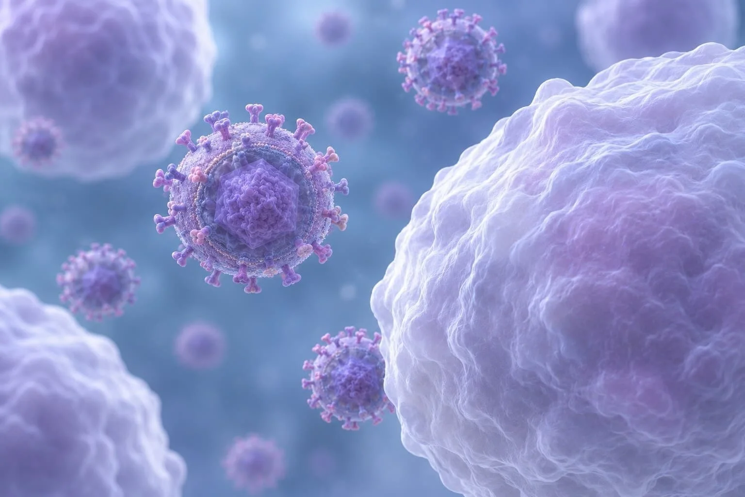
Epstein-Barr virus, or human herpesvirus 4, ranks among the most widespread viruses in people (Hatton et al., 2014; CDC, 2024a). Its DNA genome is about 170 kilobases long and holds more than 85 genes (Hatton et al., 2014). A review published in Cell in 2022 describes it as found everywhere and capable of causing cancer, with ties to several malignancies and to autoimmune disorders (Damania et al., 2022).
How many people carry the virus
Estimates run from about 90% of adults to over 95% of all people worldwide, depending on the source (Dunmire et al., 2018; Damania et al., 2022). The CDC puts the figure at about 9 in 10 adults with antibodies from a current or past infection (CDC, 2024a).
Children have lower rates. In a national survey of 8417 US residents aged 6–19, 66.5% had antibodies (Dowd et al., 2013). The share climbed from 54.1% at ages 6–8 to 82.9% at ages 18–19 (Dowd et al., 2013). Household income made a large difference, with 81.0% in the poorest quarter of families against 53.9% in the richest, but the survey could not tell whether that gap reflected more exposure, greater susceptibility or both (Dowd et al., 2013).
Spread through saliva
Saliva carries most infections (CDC, 2024a). Blood and semen can also pass the virus on, through sexual contact, transfusion or organ transplant, and one review adds breast milk to the list (CDC, 2024a; Damania et al., 2022).
Among teenagers, the main source is deep kissing (Dunmire et al., 2018). How younger children pick up the virus has not been established, although babies can be infected as soon as the antibodies they received from their mothers have faded (Balfour et al., 2015; Dowd et al., 2013). An older study of a different virus is described in how RSV spreads.
Timing matters. A newly infected person can pass the virus on for weeks, starting before any symptoms appear (CDC, 2024a). In a student cohort, thirteen people had virus in the mouth 2–36 days before they felt ill, while exercise, stress and diet showed no link with the risk of infection (Balfour et al., 2013).
From first infection to mononucleosis
Mononucleosis is a contagious illness, and Epstein-Barr virus is its most common cause, though other viruses can produce it too (CDC, 2024b). Symptoms come only after a long wait. The incubation period is about six weeks, far longer than for illnesses such as influenza (Dunmire et al., 2018). Historical observations place it at 32 to 49 days (Balfour et al., 2015).
Age at first infection
In developing countries most people meet the virus as young children, while in industrialised countries the first infection tends to come later (Damania et al., 2022). That difference is why mononucleosis is seen more often in developed countries (Balfour et al., 2015).
Childhood infection usually passes without symptoms (CDC, 2024a). Infection delayed to adolescence has been reported to end in mononucleosis in between 20% and 50% of cases (Hadinoto et al., 2008). The CDC gives a floor of at least 1 in 4 infected teenagers and young adults (CDC, 2024b).
A prospective study at the University of Minnesota screened 546 first-year students, found that 202 had no antibodies and enrolled 143 of them (Balfour et al., 2013). Over a median of 3 years, 66 caught the virus, and of these 77% developed mononucleosis, 11% had no symptoms and 12% had atypical ones (Balfour et al., 2013).
Infection was most frequent in the first year, at 26 cases per 100 person-years against 10 in the following 3 years (Balfour et al., 2013). Genes may play a part as well. In Danish registries, the rate ratio for mononucleosis was 9.3 among same-sex twins and 2.3 among parents, children and siblings (Balfour et al., 2015).
Symptoms and how long they last
Sore throat was the most common complaint among 59 students with symptoms, reported by 93%, ahead of swollen neck glands in 76% and fatigue in 66% (Balfour et al., 2013). Fever was less frequent, at 42% (Balfour et al., 2013).
Its length varies widely (Balfour et al., 2013). It lasted a median of 10 days, with an average of 17.0 days and a span of 3–66 days (Balfour et al., 2013). Fatigue and swollen glands frequently last 3 weeks or longer (Balfour et al., 2015). According to the CDC, most people with symptoms recover in 2 to 4 weeks, though tiredness can linger for weeks or even months (CDC, 2024a).
The liver is often involved. Raised liver enzymes appear in 75% of patients, usually without jaundice or pain (Balfour et al., 2015). Mononucleosis can involve the spleen, and its rare complications include splenic rupture and infarction, with rupture the one doctors fear most (Toti et al., 2023; Balfour et al., 2015). It occurs in fewer than 1% of patients (Balfour et al., 2015).
Published case reports describe 186 ruptures and 29 infarctions since 1970 (Toti et al., 2023). Timing was recorded for 145 of the ruptures, and at least 80% of cases with known timing fell within three weeks after symptoms started (Toti et al., 2023). Only 9.1% followed an injury (Toti et al., 2023). Of the reported ruptures, 4.8% were fatal, but these shares come from published case reports and cannot give the risk for an individual person with mononucleosis (Toti et al., 2023).
Why the illness feels so severe is still debated. One review describes the symptoms as likely driven by an overly strong attack by cytotoxic T cells on infected B cells (Damania et al., 2022). A study of 15 patients similarly linked the onset of symptoms to the mass killing of infected cells, rather than to the moment of infection (Hadinoto et al., 2008).
Almost all patients recover, unless a genetic defect weakens their immune response to the virus (Damania et al., 2022). Serious complications in the acute phase are rare (Balfour et al., 2015).
How the virus persists in B cells
The virus can multiply in cells lining the throat and infect B cells that pass through the mouth, although which of these cells is infected first is not fully settled (Damania et al., 2022; Wong et al., 2022). B cells are its main target because they carry CD21, the receptor the virus uses to get in (Hatton et al., 2014).
Infected young B cells then grow, pass through a germinal centre and become memory B cells, which enter the blood and keep the virus for life (Hadinoto et al., 2008). In acute mononucleosis, up to half of all memory B cells can carry it six weeks after infection (Damania et al., 2022). In 13 of 15 patients with acute illness, these infected cells then fell with an average half-life of 7.5 days, anywhere between 2.4 and 15.2 days (Hadinoto et al., 2008).
Latency programmes
The virus then goes quiet (Damania et al., 2022). During the resting phase, the viral DNA sits in the cell nucleus as a ring tethered to the host chromosomes by a viral protein called EBNA-1 (Damania et al., 2022). In quiet memory B cells the virus enters a state known as Latency 0, in which only two groups of viral RNAs, EBERs and BARTs, are made (Damania et al., 2022).
These resting cells stay silent (Hadinoto et al., 2008). They make no viral latent proteins and show no viral replication, so the immune system has little to recognise and leaves them alone, which keeps the virus out of sight (Hadinoto et al., 2008; Hatton et al., 2014). The same feature makes them harmless, which explains why lifelong infection is usually benign (Hadinoto et al., 2008).
The immune system still devotes a sizeable force to keeping the virus in check. T cells aimed at a single viral target can account for up to 2% of all CD8+ T cells in a carrier (Hatton et al., 2014). In the long run, only a few memory B cells hold the virus (Wong et al., 2022).
Reactivation and shedding
Now and then the virus switches back to making new particles, through processes that remain unclear, and the immune response usually keeps this under control (Hatton et al., 2014; Damania et al., 2022). The CDC notes that a reactivated virus can be passed to others however many years have gone by since the first infection (CDC, 2024a).
Cells lining the throat produce much of this new virus and release it into saliva (Hatton et al., 2014). After a first infection, students carried viral DNA in mouth cells for a median of 175 days, and it could still be found as long as 3 years later (Balfour et al., 2013). In the blood, the virus usually became undetectable after 200 days (Balfour et al., 2013).
Long-term carriers shed as well. In 8 healthy adults, only 8 of 173 saliva samples were negative (Hadinoto et al., 2009). Repeated rinses in two of them showed that saliva virus was fully replaced within 2 minutes, about the time between swallows, so the mouth works as a channel rather than a store (Hadinoto et al., 2009). Over months, though, levels in the same person swung by 3.5 to 5.5 logs, so whether someone counts as a high or a low shedder depends more on when they are tested than on who they are (Hadinoto et al., 2009).
Laboratory confirmation of a recent infection
Doctors usually diagnose mononucleosis from the symptoms alone (CDC, 2024b). When tests are used, they are heterophile antibody tests, antibody tests specific to the virus, or both (Dunmire et al., 2018).
Results vary with age (Balfour et al., 2015). Heterophile antibodies were found in 50 of 65 infected students (77%), and early IgM antibodies against the viral capsid in 83% (Balfour et al., 2013). Young children often lack heterophile antibodies, and about 40% of children aged 4 or under never make them (Balfour et al., 2015; CDC, 2024c). Because these antibodies can remain for a year or longer, a positive result does not always point to a recent infection (Balfour et al., 2015).
IgG antibodies against the viral capsid appear early, peak 2 to 4 weeks after onset and then stay for life, whereas antibodies against the nuclear antigen arrive slowly, 2 to 4 months after symptoms begin, and also persist (CDC, 2024c). As over 90% of adults were infected years earlier, most adults test positive for these antibodies (CDC, 2024c). About 20% of healthy people carry antibodies against the early antigen for years (CDC, 2024c).
Links between the virus and later illness
Mononucleosis has been tied to long-term consequences, above all multiple sclerosis and Hodgkin lymphoma (Balfour et al., 2015). Why these conditions are linked to a symptomatic first infection is not known (Balfour et al., 2015).
Fatigue after mononucleosis
A US study followed 301 adolescents aged 12–18 after mononucleosis confirmed by a positive monospot test and presumed to be caused by the virus (Katz et al., 2009). At 6 months, 70 of the 286 who completed a telephone screen, or 24%, had not fully recovered (Katz et al., 2009).
Chronic fatigue syndrome was diagnosed in 13% at 6 months, 7% at 12 months and 4% at 24 months (Katz et al., 2009). All 13 still affected at 24 months were girls, and most participants recovered with time (Katz et al., 2009). The authors compared these rates with a general adolescent rate of 0.2%, not with a matched group followed alongside, and concluded that mononucleosis could raise the risk (Katz et al., 2009).
Multiple sclerosis
The strongest evidence comes from a cohort of over 10 million young service members in the US, and 955 of them developed multiple sclerosis while serving (Bjornevik et al., 2022). Within that cohort, the risk of the disease rose 32-fold once people had caught Epstein-Barr virus while other viruses, cytomegalovirus among them, did not raise it even though cytomegalovirus spreads in a similar way (Bjornevik et al., 2022). The authors say no known risk factor explains the findings and put the virus forward as the main cause of multiple sclerosis (Bjornevik et al., 2022).
In the study's blood samples, a marker of nerve damage, neurofilament light chain, increased only after seroconversion, the point at which antibodies to the virus appear (Bjornevik et al., 2022). Over 90% of adults carry the virus (Hatton et al., 2014). A relative increase of this size therefore does not mean that a given carrier is likely to develop multiple sclerosis. How the immune system can turn on the body's own tissues is explained in what autoimmune disease is.
Cancers associated with the virus
In 1997 the cancer agency of the WHO, IARC, put the virus in its top group of carcinogens because of its causal links with nasopharyngeal carcinoma, endemic Burkitt lymphoma and Hodgkin lymphoma (Wong et al., 2022). One estimate attributes about 84.6% of new nasopharyngeal carcinoma cases to the virus (Wong et al., 2022). In parts of southern China this cancer reaches 20–50 cases per 100,000 people (Damania et al., 2022).
More than 90% of endemic Burkitt lymphomas, often jaw tumours in children, contain the virus (Damania et al., 2022). The share of Hodgkin lymphoma linked to it varies by region, from 74% in Africa to 32% in North America and 29% in Oceania (Wong et al., 2022). After mononucleosis, the risk of virus-positive Hodgkin lymphoma is about four times higher (Damania et al., 2022). Additional factors can tip the balance towards cancer, such as genetic changes, a weakened immune system, HIV infection or a diet high in salted or preserved fish (Damania et al., 2022).
Stomach cancer is less clear-cut (Wong et al., 2022). About 10% of stomach cancers carry the virus, but a causal role there remains unproven (Damania et al., 2022; Wong et al., 2022). In 2020, six cancer types linked to the virus, stomach cancer among them, made up an estimated 239,700–357,900 new diagnoses, with 137,900–208,700 deaths, or about 1.3–1.9% of the world's cancers (Wong et al., 2022). The authors note that their ranges may be too wide in places, while rarer cancers left out of the count make the total conservative (Wong et al., 2022).
Measure | Group studied | Result |
Antibodies to the virus | 8417 US residents aged 6–19 | 66.5% positive |
Mononucleosis after a first infection | 66 infected students | 77% |
Length of illness | 59 students with symptoms | Median 10 days |
Virus in mouth cells | 66 infected students | Median 175 days |
Chronic fatigue syndrome at 6 months | 301 adolescents after mononucleosis | 13% |
Multiple sclerosis | Service members in a cohort of over 10 million | Risk 32-fold higher after infection |
Note: the rows come from different populations and study designs (Dowd et al., 2013; Balfour et al., 2013; Katz et al., 2009; Bjornevik et al., 2022). They describe groups rather than the chance for any one person, and the multiple sclerosis figure is a relative increase, not an absolute risk.
What is established about Epstein-Barr virus and what remains uncertain
Some points are settled. Most people carry the virus, often from childhood, and it lives on quietly in memory B cells (CDC, 2024a; Damania et al., 2022). Infection delayed until the late teens is often followed by mononucleosis, and patients almost always recover (Balfour et al., 2013; Damania et al., 2022).
Among 66 university students who caught the virus, 77% developed mononucleosis, and the illness lasted a median of 10 days in those with symptoms (Balfour et al., 2013).
Much remains open. Researchers still do not know which viral and immune factors decide whether an infection stays silent, causes mononucleosis or leads to cancer (Hatton et al., 2014). Its complicated life cycle has so far stood in the way of an effective vaccine (Hatton et al., 2014; Dunmire et al., 2018). The CDC states that there is no vaccine and no specific treatment (CDC, 2024a).
The long-term links rest mainly on observational data. The multiple sclerosis findings point strongly to the virus but come from one cohort of service members (Bjornevik et al., 2022). The fatigue study had no matched comparison group, and the splenic rupture figures come from published cases rather than population counts (Katz et al., 2009; Toti et al., 2023).
Frequently asked questions
Can a person get mono more than once?
Repeat illness seems uncommon. After a first infection the virus stays in the body and may reactivate (CDC, 2024a). In students followed after infection, there were 2 reported clinical relapses during 125.5 person-years, and only one came with a rise in viral load (Balfour et al., 2013). A mono-like illness can also come from other viruses (CDC, 2024b).
Is there a vaccine against Epstein-Barr virus?
No. The CDC states that no vaccine protects against Epstein-Barr virus infection (CDC, 2024a). Researchers report that several attempts have not produced an effective preventive vaccine, partly because the viral life cycle is so complex (Dunmire et al., 2018; Hatton et al., 2014). There is also no specific approved treatment for mononucleosis (Balfour et al., 2015).
How long does the virus stay in saliva after mono?
Usually for months. In students followed after a first infection, viral DNA stayed in mouth cells for a median of 175 days, and in some it was still found 3 years later (Balfour et al., 2013). A smaller study found that shedding was still slowly declining a year after the acute illness (Hadinoto et al., 2008).
Why is mono called the kissing disease?
Kissing is the best-documented route among young people. Exposure to saliva during deep kissing is identified as the main source of a first infection in adolescents (Dunmire et al., 2018). In a cohort of university students, kissing was a significant risk factor (Balfour et al., 2013). The virus can also spread through blood, semen and organ transplants (CDC, 2024a).
Does everyone with the virus develop mono?
No. Most people are infected in childhood and never have symptoms (CDC, 2024a). Mono is more likely when the first infection comes in the teens or later, and the CDC says at least 1 in 4 infected teenagers and young adults develop it, while one student cohort found 77% (CDC, 2024b; Balfour et al., 2013).
Sources
- Damania B, Kenney SC, Raab-Traub N. Epstein-Barr virus: biology and clinical disease. Cell. 2022;185(20):3652–3670. DOI 10.1016/j.cell.2022.08.026.
- Hatton OL, Harris-Arnold A, Schaffert S, Krams SM, Martinez OM. The interplay between Epstein-Barr virus and B lymphocytes: implications for infection, immunity, and disease. Immunologic Research. 2014;58(2–3):268–276. DOI 10.1007/s12026-014-8496-1.
- Hadinoto V, Shapiro M, Greenough TC, Sullivan JL, Luzuriaga K, Thorley-Lawson DA. On the dynamics of acute EBV infection and the pathogenesis of infectious mononucleosis. Blood. 2008;111(3):1420–1427. DOI 10.1182/blood-2007-06-093278.
- CDC (Centers for Disease Control and Prevention). About Epstein-Barr Virus (EBV). 2024a, page dated 9 May 2024. https://cdc.gov/epstein-barr/about/index.html
- Dowd JB, Palermo T, Brite J, McDade TW, Aiello A. Seroprevalence of Epstein-Barr virus infection in U.S. children ages 6–19, 2003–2010. PLoS ONE. 2013;8(5):e64921. DOI 10.1371/journal.pone.0064921.
- Balfour HH Jr, Odumade OA, Schmeling DO, Mullan BD, Ed JA, Knight JA, Vezina HE, Thomas W, Hogquist KA. Behavioral, virologic, and immunologic factors associated with acquisition and severity of primary Epstein-Barr virus infection in university students. Journal of Infectious Diseases. 2013;207(1):80–88. DOI 10.1093/infdis/jis646.
- Hadinoto V, Shapiro M, Sun CC, Thorley-Lawson DA. The dynamics of EBV shedding implicate a central role for epithelial cells in amplifying viral output. PLoS Pathogens. 2009;5(7):e1000496. DOI 10.1371/journal.ppat.1000496.
- Balfour HH Jr, Dunmire SK, Hogquist KA. Infectious mononucleosis. Clinical & Translational Immunology. 2015;4(2):e33. DOI 10.1038/cti.2015.1.
- Dunmire SK, Verghese PS, Balfour HH Jr. Primary Epstein-Barr virus infection. Journal of Clinical Virology. 2018;102:84–92. DOI 10.1016/j.jcv.2018.03.001.
- Katz BZ, Shiraishi Y, Mears CJ, Binns HJ, Taylor R. Chronic fatigue syndrome after infectious mononucleosis in adolescents. Pediatrics. 2009;124(1):189–193. DOI 10.1542/peds.2008-1879.
- Bjornevik K, Cortese M, Healy BC, Kuhle J, Mina MJ, Leng Y, Elledge SJ, Niebuhr DW, Scher AI, Munger KL, Ascherio A. Longitudinal analysis reveals high prevalence of Epstein-Barr virus associated with multiple sclerosis. Science. 2022;375(6578):296–301. DOI 10.1126/science.abj8222.
- Wong Y, Meehan MT, Burrows SR, Doolan DL, Miles JJ. Estimating the global burden of Epstein-Barr virus-related cancers. Journal of Cancer Research and Clinical Oncology. 2022;148(1):31–46. DOI 10.1007/s00432-021-03824-y.
- Toti JMA, Gatti B, Hunjan I, Kottanattu L, Faré PB, Renzi S, Bianchetti MG, Milani GP, Lava SAG, Camozzi P. Splenic rupture or infarction associated with Epstein-Barr virus infectious mononucleosis: a systematic literature review. Swiss Medical Weekly. 2023;153:40081. DOI 10.57187/smw.2023.40081.
- CDC (Centers for Disease Control and Prevention). About Infectious Mononucleosis (Mono). 2024b, page dated 9 May 2024. https://cdc.gov/epstein-barr/about/mononucleosis.html
- CDC (Centers for Disease Control and Prevention). Laboratory Testing for Epstein-Barr Virus (EBV). 2024c, page dated 10 April 2024. https://cdc.gov/epstein-barr/php/laboratories/index.html

Tomas Verren — author
Tomas Verren has a background in biomedical sciences and population health, with academic training in human biology, epidemiology, study design and the interpretation of health data. After completing his postgraduate training, he moved into research communicat...
This article is for general information and is not medical advice. It was reviewed for accuracy by a qualified clinician; decisions about your health should be made with your own doctor.
Related
What autoimmune disease is and why the immune system attacks the body

How RSV spreads: lessons from a 1981 transmission study
How staph infections start on the skin and reach the blood

Polio vaccines in 1960: what the debate got right and wrong

Swimmer's ear and why water in the ear canal leads to infection
Pseudomonas aeruginosa and why this bacterium is so hard to treat
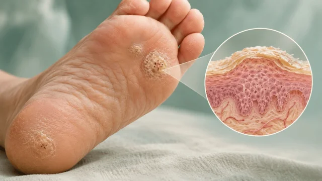
How HPV causes plantar warts on the soles of the feet