




Skin tumours in dogs and cats: what pathology data show
Skin tumours are a major part of small-animal diagnostic pathology because a visible lump can represent anything from a benign growth or cyst to a malignant neoplasm. Goldschmidt and Shofer's 1992 book drew on a very large University of Pennsylvania pathology archive to describe the types, microscopic features and demographic patterns of canine and feline skin tumours.
The book's headline figures need careful interpretation. Between 1985 and 1990 the laboratory received 65,000 dog specimens and 13,250 cat specimens, with skin tumours and tumour-like lesions accounting for 45% and 24% respectively. Those percentages describe submitted pathology specimens, not the incidence of skin cancer in the general dog or cat population.
Why a pathology archive was valuable
The 1992 volume was built from specimens submitted for surgical pathological evaluation to the Pathology Laboratory at the University of Pennsylvania School of Veterinary Medicine. That made it possible to assemble one of the largest structured datasets of canine and feline skin lesions available at the time.
A pathology archive offers something a simple case series cannot: repeated microscopic diagnoses made across many years, animals and lesion types. The book used those records to organize tumour categories and to describe age, sex, anatomical-site and breed predilections. For clinicians and pathologists, those patterns can help frame differential diagnoses, but they do not replace examination of the individual mass.
What the 45% and 24% figures actually mean
The abstract reports that 65,000 dog specimens and 13,250 cat specimens were received between 1985 and 1990, and that skin tumours and tumour-like lesions represented 45% of canine submissions and 24% of feline submissions. The denominator is therefore the material sent to a veterinary pathology service, not all dogs or cats cared for in the community.
That distinction is crucial. Animals whose tissue is submitted for histopathology have already been selected because a veterinarian or owner considered a lesion worth investigating. Referral patterns, surgical practice, breed distribution, geography and the kinds of cases seen by the institution all influence what reaches the laboratory. The figures are best read as the burden of cutaneous lesions within this pathology workload rather than as lifetime risk or population prevalence.
The comparison is between species within the same submission archive
The 45% value belongs to dog submissions and the 24% value to cat submissions. They are not before-and-after measurements, treatment effects or malignancy rates. Nor do they mean that 45% of dogs or 24% of cats developed a tumour. The comparison reflects how frequently skin tumours and tumour-like lesions appeared among the two species' submitted specimens during the stated period.
How to interpret the source data
Species | Total pathology specimens | Skin tumours / tumour-like lesions | Interpretation |
Dogs | 65,000 | 45% of submissions | Pathology-laboratory proportion, not population incidence |
Cats | 13,250 | 24% of submissions | Pathology-laboratory proportion, not population incidence |
Why histopathology matters for skin masses
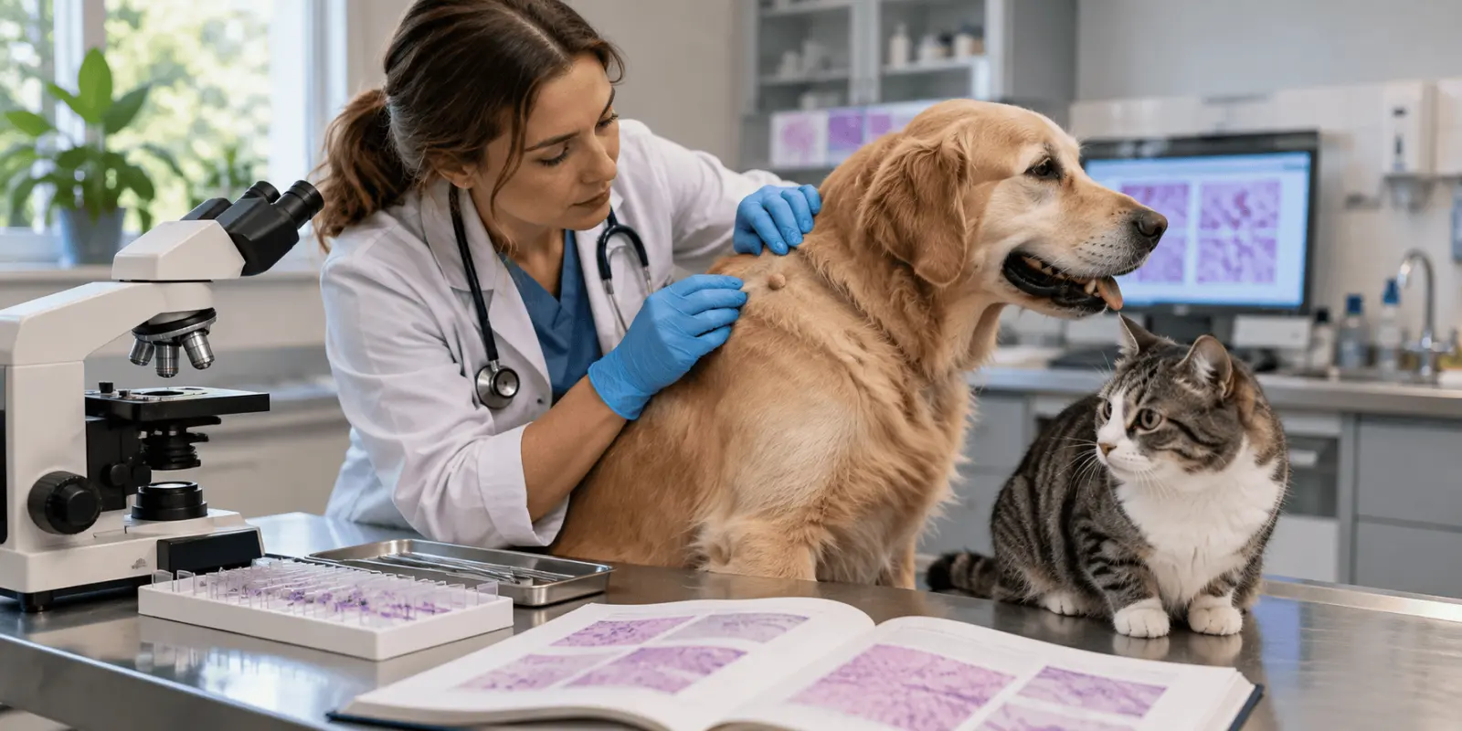
A skin mass cannot be classified reliably by appearance alone. Benign and malignant lesions can overlap in size, colour, firmness and growth pattern, and inflammatory or cystic lesions can mimic tumours. Current veterinary references therefore emphasize cytology and histopathology as complementary diagnostic tools. Fine-needle aspiration can often provide an early cellular assessment, while biopsy allows the architecture of the tissue to be examined.
Histopathology can establish tumour type, describe cellular differentiation and, for selected tumours, provide grade or other features linked to biological behaviour. For surgically removed masses it can also assess whether tumour cells extend to the excision margin. These details influence whether observation, further surgery, staging, radiation therapy, chemotherapy or other management may be considered.
The diagnostic value of a pathology result also depends on the question being asked. Cytology can be useful for an initial assessment of a mass, particularly when cells can be obtained easily, while histopathology provides information about tissue architecture that a cellular sample cannot show. That distinction matters when a veterinarian needs to separate closely related tumour types, assess patterns of invasion or evaluate an excised lesion. For some neoplasms, microscopic grading adds prognostic information, while for others the main task is accurate classification. Pathology therefore works best when the microscopic findings are interpreted together with the animal’s age, breed, lesion site, clinical appearance and treatment history. The 1992 book was built around this same principle of linking morphology with signalment and anatomical location, even though modern diagnostic systems now use more refined criteria for several tumour groups.
A visible lump is a clinical finding; microscopic classification determines what kind of lesion it actually is.
Skin tumours are not a single disease
The skin contains epithelial cells, hair follicles, glands, connective tissue, blood vessels, melanocytes and immune cells, so cutaneous neoplasia includes many biologically different entities. Goldschmidt and Shofer's book was valuable precisely because it organized a broad range of these lesions and paired morphology with signalment and site information.
Later veterinary pathology classifications continued this work. The World Health Organization's second series on tumours of domestic animals included dedicated classifications for epithelial and melanocytic skin tumours and for mesenchymal tumours of skin and soft tissue. Modern pathology texts likewise separate epithelial, melanocytic, mesenchymal and round-cell or haematopoietic neoplasms because diagnosis and prognosis depend on cell lineage and tumour subtype.
Examples differ between dogs and cats
The mix of common lesions is not identical across species or regions. A large UK study of more than 9,000 feline cutaneous tumours found basal cell tumours, fibrosarcomas, squamous cell carcinomas and mast cell tumours among the most frequent diagnoses. A Japanese retrospective study of 1,435 canine cutaneous tumours reported soft-tissue sarcomas, mast cell tumours, lipomas, hair-follicle tumours and benign sebaceous tumours among the leading categories.
Those later studies provide comparative context rather than replacing the 1992 book's dataset. They come from different countries, periods, laboratory populations and inclusion criteria. Their value is comparative: they show that the distribution of skin-tumour diagnoses varies with the population and methodology, which reinforces why pathology datasets must always be interpreted in context.
Differences between pathology datasets can arise even when laboratories use similar diagnostic terminology. Referral hospitals, university laboratories and first-opinion practices do not necessarily receive the same mix of cases, and breed distributions can vary substantially between countries. Changes in diagnostic criteria over time can also affect how older and newer tumour categories are compared. For that reason, frequency rankings are most informative within the population from which they were generated. The UK feline and Japanese canine studies are useful because they show how large modern datasets can describe species-specific patterns, but they should be interpreted alongside their own sampling frames rather than treated as direct extensions of the Pennsylvania archive. This is also why the large numbers in Goldschmidt and Shofer’s book are valuable descriptively without being population-incidence estimates.
Signalment can guide suspicion but not make the diagnosis
The 1992 book catalogued age, sex, site and breed predilections for specific tumours. Such patterns are clinically useful because some tumour types appear disproportionately in certain ages, breeds or anatomical locations. However, a predilection changes probability rather than proving identity. A mass in a breed associated with a particular neoplasm can still represent a completely different lesion.
Large later datasets support the same principle. The Japanese canine study found age and breed associations for several cutaneous tumour types, while the UK feline study identified breed differences for some diagnoses. These epidemiological patterns can help prioritize differentials, but microscopic evaluation remains necessary when the nature of the lesion will affect treatment or prognosis.
Mast cell tumours illustrate why classification evolves
Canine mast cell tumours are a useful example of how veterinary oncology has changed since the early 1990s. They are often diagnosable cytologically, but biopsy and histopathological grading are important for prognosis and treatment planning. Older three-tier grading systems were widely used, yet studies demonstrated substantial disagreement between pathologists, prompting development of a two-tier system intended to improve reproducibility.
More recent consensus work has refined definitions for cutaneous, subcutaneous and other mast cell neoplasms and emphasizes combining histological findings with staging and clinical information. This does not diminish the historical value of Goldschmidt and Shofer's book. Instead, it shows how large descriptive pathology collections provide a foundation that later studies can standardize and test more rigorously.
What the 1992 book can and cannot tell us today
The book remains valuable for historical pathology, terminology, morphology and the epidemiological patterns present in a major referral laboratory at the end of the 1980s. Its large case base gives a detailed snapshot of what veterinary pathologists were diagnosing and how tumours were distributed by species, age, sex, breed and body site.
It cannot, however, provide a current population incidence of skin cancer in dogs or cats. Diagnostic criteria have evolved, breed populations have changed, referral pathways differ between countries and decades, and some tumour entities are now classified or graded differently. The source also combines tumours with tumour-like lesions in its headline submission percentages, so those percentages do not represent malignant-tumour rates.
The referral nature of the original dataset is particularly important when interpreting percentages. A pathology service receives animals because a lesion has already been noticed and sampled, so its case mix differs from the wider population of dogs and cats. Frequencies within that archive describe what reached the laboratory, not the probability that any randomly selected animal will develop a particular tumour. Modern epidemiological studies need defined source populations and denominators before they can estimate incidence or risk. The book remains valuable because it documents diagnostic patterns in a very large pathology series, but those patterns should not be converted into population-wide cancer rates. This distinction is essential when historical pathology archives are used to inform present-day clinical decisions or epidemiological comparisons.
For a present-day animal with a new or changing skin mass, the historical statistics are background information rather than a diagnosis. Veterinary assessment is needed to decide whether cytology, biopsy or excision is appropriate and how pathology findings should be integrated with the animal's overall health and tumour location.
Why the book still matters in veterinary pathology
Skin Tumors of the Dog and Cat helped turn a large pathology workload into a structured reference. Its central contribution was not a single percentage but the systematic linkage of microscopic diagnosis with clinical descriptors and population patterns. That approach remains fundamental to veterinary tumour pathology: classify the lesion accurately, describe its behaviour-related features and interpret epidemiological patterns without confusing a referral dataset with the wider pet population.
Frequently asked questions
Does the 45% figure mean 45% of dogs develop skin tumours?
No. The 45% figure refers to the proportion of dog pathology specimens submitted to one university laboratory between 1985 and 1990 that were classified as skin tumours or tumour-like lesions. It is not a population incidence, lifetime risk or estimate for all dogs.
What was the source of the data in Goldschmidt and Shofer's book?
The book was based on specimens submitted for surgical pathological evaluation to the University of Pennsylvania School of Veterinary Medicine. The abstract reports 65,000 dog specimens and 13,250 cat specimens received between 1985 and 1990, with detailed tumour descriptions compiled from that archive.
Can a veterinarian identify a skin tumour just by looking at it?
Usually not with enough certainty for treatment planning. Different benign, malignant, inflammatory and cystic lesions can look similar. Fine-needle aspiration may provide a rapid cytologic assessment, while biopsy and histopathology are often needed to establish tumour type, evaluate tissue architecture and assess features such as surgical margins.
Are the most common skin tumours the same in dogs and cats?
No. The distribution differs by species and also varies among studies, regions and referral populations. Later feline and canine retrospective studies report different leading tumour categories. Frequency data from one pathology archive therefore do not automatically apply to another country, period or patient population.
Why is histological grading important for some skin tumours?
For selected neoplasms, microscopic grade helps estimate biological behaviour and prognosis. Canine cutaneous mast cell tumours are a well-known example: grading systems assess cellular and mitotic features, and newer schemes were developed partly to improve agreement between pathologists. Grade is interpreted alongside staging and clinical information.
Sources
- Goldschmidt MH, Shofer FS. Skin Tumors of the Dog and Cat. Oxford: Pergamon Press; 1992. 316 pp. ISBN 9780080408231.
- Villalobos AE. Overview of Tumors of the Skin and Soft Tissues in Animals. Merck Veterinary Manual. Updated 2024.
- Ho NT, Smith KC, Dobromylskyj MJ. Retrospective study of more than 9000 feline cutaneous tumours in the UK: 2006–2013. Journal of Feline Medicine and Surgery. 2018;20(2):128–134. doi:10.1177/1098612X17699477.
- Kok MK, Chambers JK, Tsuboi M, Nishimura R, Tsujimoto H, Uchida K, Nakayama H. Retrospective study of canine cutaneous tumors in Japan, 2008–2017. Journal of Veterinary Medical Science. 2019;81(8):1133–1143.
- Kiupel M, Webster JD, Bailey KL, et al. Proposal of a 2-Tier Histologic Grading System for Canine Cutaneous Mast Cell Tumors to More Accurately Predict Biological Behavior. Veterinary Pathology. 2011;48(1):147–155. doi:10.1177/0300985810386469.
- Willmann M, Yuzbasiyan-Gurkan V, Marconato L, et al. Proposed Diagnostic Criteria and Classification of Canine Mast Cell Neoplasms: A Consensus Proposal. Frontiers in Veterinary Science. 2021;8:755258.
- Goldschmidt MH, Dunstan RW, Stannard AA, von Tscharner C, Walder EJ, Yager JA. Histological Classification of Epithelial and Melanocytic Tumors of the Skin of Domestic Animals. WHO International Histological Classification of Tumors of Domestic Animals, Second Series, Vol III. Washington, DC; 1998.
- Roccabianca P, et al. Review of diagnostic histologic features of cutaneous round cell neoplasms in dogs. Veterinary Pathology. 2022.

Imogen Farrant — author
Imogen Farrant is an animal health and welfare writer with a background in animal science and a particular interest in companion animal welfare. Her work focuses on translating veterinary and animal-welfare research into clear, accessible information for pet o...
Related
Hereditary dwarfism in beef cattle: genetics and calf outcomes

Biting midges: biology, disease transmission and control
Blackflies: biology, life cycle, ecology and disease-vector roles

Insect pheromones and chemical communication in social insects

Animal Translocation Stress: Conservation Risks and Management